0213
Silent MRF : Quantitative scan with reduced noise using the Magnetic Resonance Fingerprinting (MRF) framework1Radiology, Case Western Reserve University, Cleveland, OH, United States
Synopsis
Because MRF provides a flexible framework without any requirement for constant repetition times or gradient amplitudes, we propose to use arbitrary gradient waveforms and randomized TR times to reduce the acoustic noise, while still providing quantitative 3D T1 and T2 values.
Introduction
MRF scans have shown high scan efficiency and repeatability on quantifying tissue properties, such as T1 and T2 values(1,2). However, because fast switching gradients for the slice encoding and spiral readout are typically applied, the acoustic noise generated from the MRF scan is louder than the clinical routine scans, with an average sound pressure level (aSPL) of 88.4 dB and maximum SPL (mSPL) of 90.3 dB from an MRF scan, as compared to aSPL of 84.1 dB and mSPL of 85.8 dB for a diffusion scan and aSPL of 81.4 dB and mSPL of 83.5 dB for a TSE scan. Because MRF provides a flexible framework without any requirement for constant repetition times or gradient amplitudes, we propose to use arbitrary gradient waveforms and randomized TR times to reduce the acoustic noise, while still providing quantitative 3D T1 and T2 values.Methods
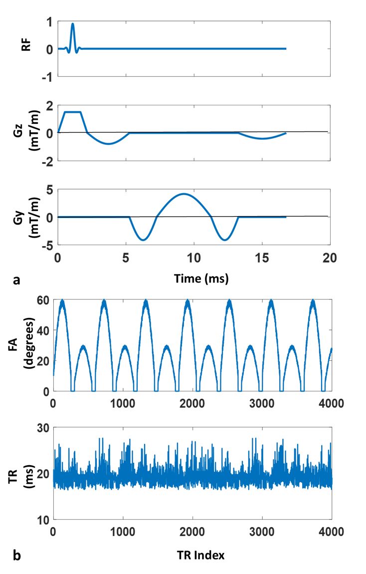
Here we propose to use three steps to reduce the acoustic noise: first, half-sine waveforms with slow slew rates were used for the gradients from all three axes, as shown in Figure 1a. Such gradient shapes have been shown to reduce the acoustic noise by slowing down the slew rate(3,4). To ensure a consistent slice encoding and good slice profile, a trapezoidal slice selection gradient with low amplitude and slow slew rate was used in each TR. Because the slice selection gradients had varying durations, and the ramp up/down time was also used for excitation, VERSE pulse(5) was designed in each TR based on the varying gradient shape. Second, randomized timing, including RF duration, slice encoding time, readout time and total TR time, was applied so that the noise spectrum is distributed and doesn’t excite any acoustic resonances in the scanner. Third, a 3D-MRF scan with slab selective excitation was implemented to provide a volumetric coverage. The amplitude of the slice selective gradient is also much lower when a large slab is excited, so that the noise from the gradient can be substantially reduced.
A phantom and an in vivo scan with a FOV of 300x300x80 mm3 and image resolution of 1.2x1.2x5 mm3 were performed in a Siemens 3T Skyra system. For each partition, 4000 time-points were acquired and the FA and TR patterns were shown in Figure 1b. The acquisition time was 76 seconds per partition. The RF pulse at each TR was designed to have the same slice profile as a sinc pulse with a duration of 2000 us and time bandwidth product of 8. As shown in Figure 1a, a balanced and smoothly varying waveform was applied in Gy in each TR. The resulting k-space trajectory was similar to the radial trajectory, but with varying step size along the radial direction. Only one such radial trajectory was used to reconstruct an image in each TR, which leads to an undersampling factor of around 400. This gradient with randomized duration was rotated at a golden-angle from one TR to the next. At the end of each TR, a spoiling gradient with a half-sine shape was applied to achieve a 4-pi dephasing across the partition thickness. After the acquisition, the reconstruction and pattern recognition steps were similar to the conventional MRF methods and the MRF-Music scan(6). The aSPL and mSPL values of all scans were measured in the scanner room using an iPhone app (Toolbox Pro). The measurement time for each scan was 40 seconds.
Results
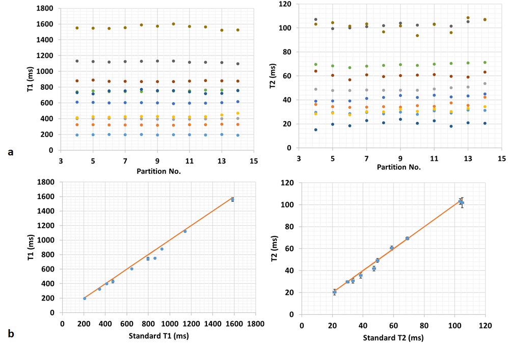
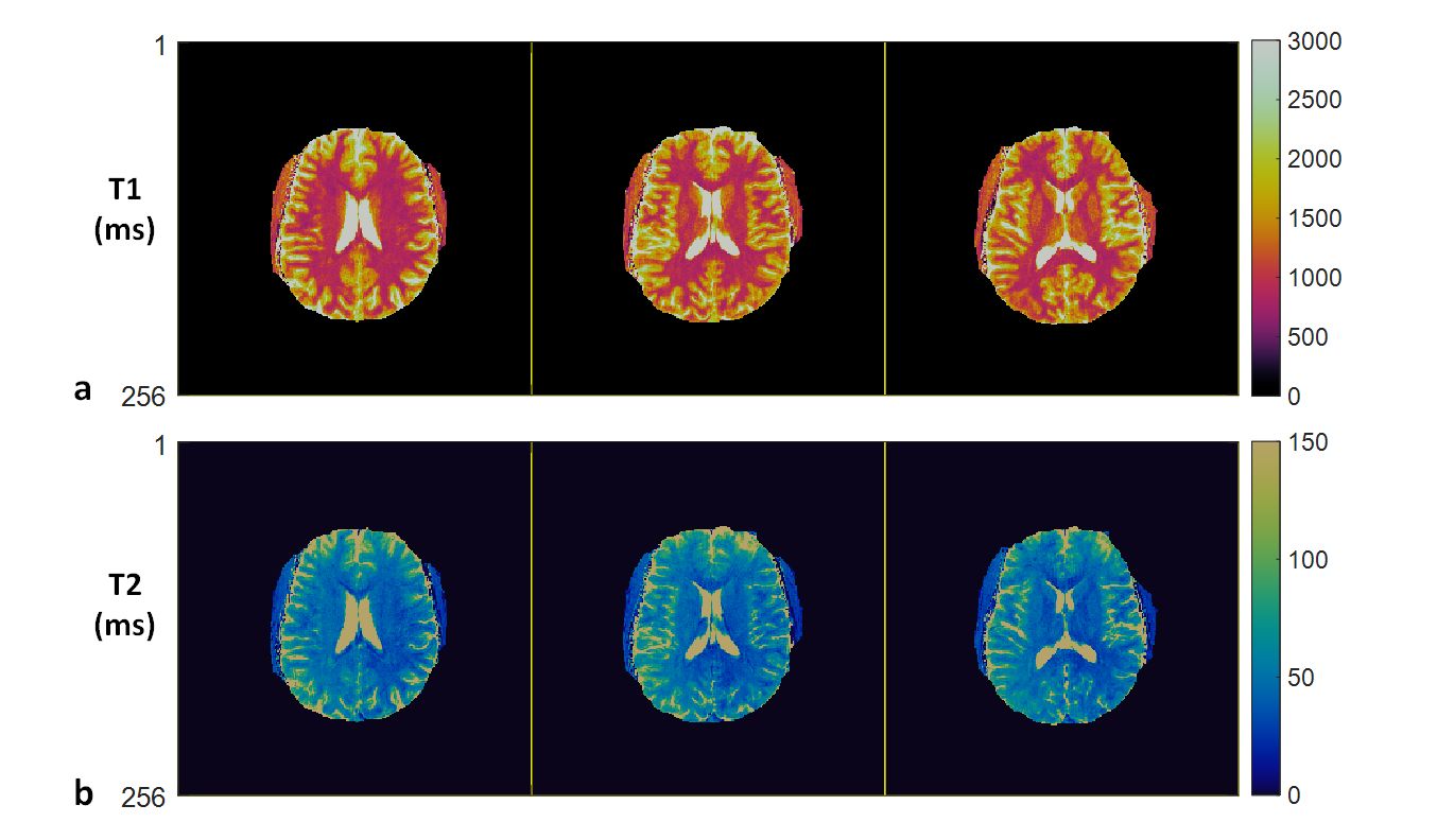
The aSPL and mSPL from the 3D silent-MRF scan is 69.8 dB and 72.3 dB, respectively, which is very close to the Siemens Quiet T2-weighted scan, with an aSPL of 70.1 dB and mSPL of 71.9 dB. As a comparison, the aSPL and mSPL from the quietest Siemens Quiet scan (T1w-PETRA) is 56.6 dB and 58.3 dB, respectively, and the ambient aSPL and mSPL from the scanner room without any scanning is 56.2 dB and 58.0 dB, respectively. Figures 2 shows the phantom results from the central 11 partitions that covers the physical length of the phantoms. The T1 and T2 values from all partitions are consistent and their average values are in good agreement with the values measured from the standard measurements. Figure 3 shows the T1 and T2 maps from different partitions of the in vivo study.Discussion
This study demonstrates a proof-of-principle implementation of the silent-MRF scan. The noise level can be further reduced by using non-selective 3D scan with no slice encoding gradient or using continuous gradients with minimum gradient steps(7,8). Given the large flexibility of the sequence design from the MRF framework, this sequence will be further optimized and advanced reconstruction methods will be incorporated to achieve better sampling efficiency and higher acceleration, without increasing the acoustic noise.Acknowledgements
The authors would like to acknowledge funding from Siemens Healthcare and NIH grants NIH 1R01EB016728-01A1 and NIH 5R01EB017219-02References
1. Ma D, Gulani V, Seiberlich N, Liu K, Sunshine JL, Duerk JL, Griswold MA. Magnetic Resonance Fingerprinting. Nature [Internet] 2013;495:187–192. doi: 10.1038/nature11971.
2. Jiang Y, Ma D, Seiberlich N, Gulani V, Griswold M a. MR fingerprinting using fast imaging with steady state precession (FISP) with spiral readout. Magn. Reson. Med. [Internet] 2014;0:n/a-n/a. doi: 10.1002/mrm.25559.
3. Hennel F. Fast spin echo and fast gradient echo MRI with low acoustic noise. J. Magn. Reson. Imaging 2001;13:960–966. doi: 10.1002/jmri.1138.
4. Heismann B, Ott M, Grodzki D. Sequence-based acoustic noise reduction of clinical MRI scans. Magn. Reson. Med. 2015;73:1104–1109. doi: 10.1002/mrm.25229.
5. Pauly J, Nishimura D, Macovski A. A k-space analysis of small-tip-angle excitation. J. Magn. Reson. 1989;81:43–56. doi: 10.1016/j.jmr.2011.09.023.
6. Ma D, Pierre EY, Jiang Y, Schluchter MD, Setsompop K, Gulani V, Griswold MA. Music-based magnetic resonance fingerprinting to improve patient comfort during MRI examinations. Magn. Reson. Med. [Internet] 2015. doi: 10.1002/mrm.25818.
7. Grodzki DM, Jakob PM, Heismann B. Ultrashort Echo Time Imaging Using Pointwise Encoding Time Reduction With Radial Acquisition ( PETRA ). Magn. Reson. Med. 2012;67:510–518. doi: 10.1002/mrm.23017.
8. Alibek S, Vogel M, Sun W, Winkler D, Baker CA, Burke M, Gloger H. Acoustic noise reduction in MRI using Silent Scan: An initial experience. Diagnostic Interv. Radiol. 2014;20:360–363. doi: 10.5152/dir.2014.13458.
Figures