0207
Maxwell-compensated waveform design for asymmetric diffusion encoding1Clinical Sciences, Lund, Lund University, Lund, Sweden, 2Random Walk Imaging AB, Lund, Sweden
Synopsis
Asymmetric gradient waveforms enable efficient diffusion encoding but suffer from signal bias and image artifacts due to Maxwell terms (concomitant fields). We propose a novel method for generating "Maxwell-compensated" waveforms, and we demonstrate that such waveforms retain superior efficiency and exhibit negligible effects due to concomitant fields.
Introduction
Diffusion encoding gradient waveforms that go beyond the pulsed gradient regime can be made asymmetric around the refocusing pulse(s) to achieve time-efficient b-tensor encoding1, reduced eddy-currents2, reduced sensitivity to motion3 etc. However, unlike symmetric waveforms, asymmetric designs are sensitive to Maxwell terms (concomitant fields) which may cause significant artifacts, signal bias, and model parameter error4-6. Here, we describe a novel method to generate "Maxwell-compensated" waveforms that yield negligible effects due to Maxwell terms, and we validate the approach in simulations and measurements.Theory
The Maxwell equations dictate that linear magnetic field gradients, such as those used for diffusion sensitization, are accompanied by spatially dependent concomitant gradients. In a spin-echo sequence with the prescribed encoding gradient $$$\mathbf{G}(t)=[G_x(t)~G_y(t)~G_z(t)]^\text{T}$$$, these fields can lead to a non-zero residual moment at readout, described by a wave vector5
$$\mathbf{k}=\frac{\gamma}{2\pi}\left(\int_{\text{P1}}^{}\mathbf{E}(t)\text{d}t-\int_{\text{P2}}^{}\mathbf{E}(t)\text{d}t\right),\quad\text{Eq.1}$$
where integration is performed over periods before/after the refocusing pulse (P1/P2), and the true gradient $$$\mathbf{E}$$$ at location $$$\mathbf{r}=[x~y~z]^\text{T}$$$ is5
$$\mathbf{E}(t,\mathbf{r})=\mathbf{G}(t)+\frac{1}{4B_0}\begin{vmatrix}G^2_z(t)&0&-2G_x(t)G_z(t)\\0&G^2_z(t)&-2G_y(t)G_z(t)\\-2G_x(t)G_z(t)&-2G_y(t)G_z(t)&4G_x^2(t)+4G_y^2(t)\end{vmatrix}\mathbf{r}. \quad\text{Eq.2}$$
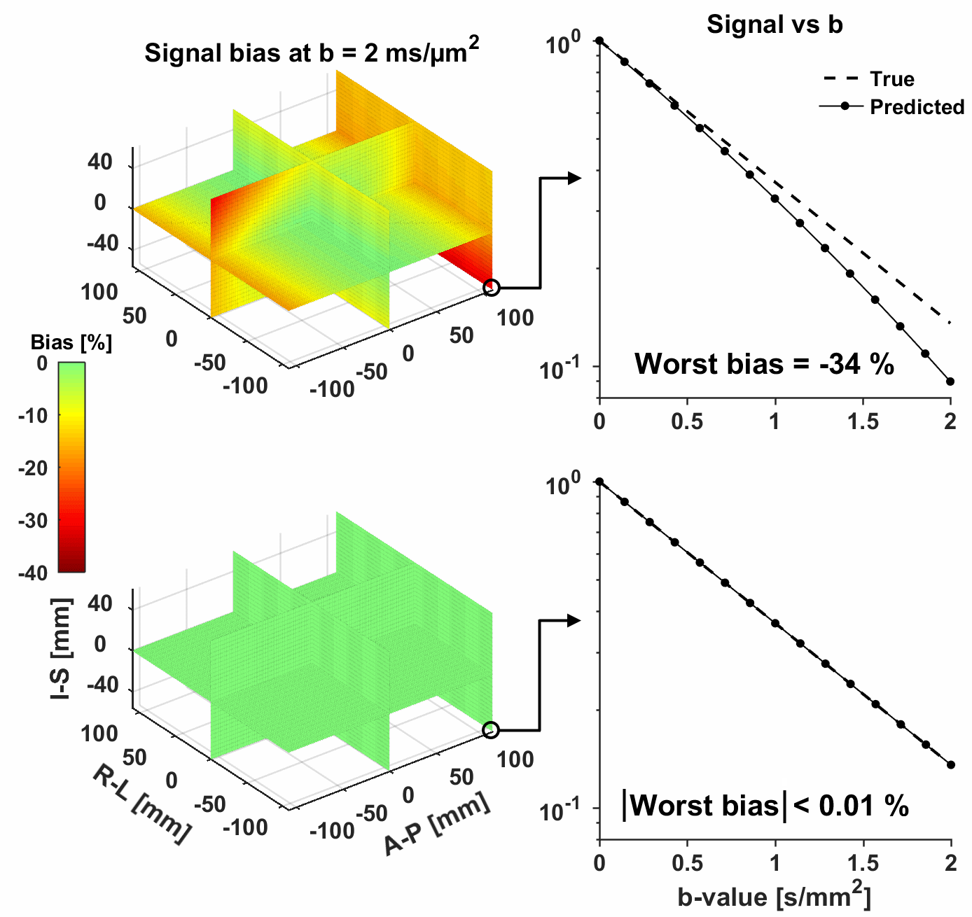
Whenever $$$\mathbf{k}\neq0$$$, data may be corrupted by artifacts and signal attenuation due to through-plane dephasing and T2* decay4. We use the attenuation factor $$$(0\leq\text{AF}\leq1)$$$ as a metric of the signal bias caused by Maxwell terms, according to
$$\text{AF}=\text{AF}_\text{slice}\cdot\text{AF}_\text{phase}=\underbrace{|\text{sinc}(\mathbf{n}_\text{slice}\cdot\mathbf{k}\cdot\text{ST})|}_{\text{AF}_\text{slice}}\cdot\underbrace{\exp(-|\mathbf{n}_\text{phase}\cdot\mathbf{k}|\cdot{\Delta}t/({\Delta}k{\cdot}T_2^*))}_{\text{AF}_\text{phase}},\quad\text{Eq.3}$$
for a rectangular slice profile where $$$\mathbf{n}_\text{slice}$$$ is the normal unit vector, $$$\mathbf{n}_\text{phase}$$$ is a unit vector along the phase encoding direction, $$$\text{ST}$$$ is the slice thickness, $$${\Delta}k$$$ is the distance between k-space lines, and $$${\Delta}t$$$ is the echo spacing. Note that $$${\Delta}k=N/FOV_\text{p}$$$, where $$$N$$$ is the in-plane acceleration factor and $$$FOV_\text{p}$$$ is the field of view along $$$\mathbf{n}_\text{phase}$$$.
Methods
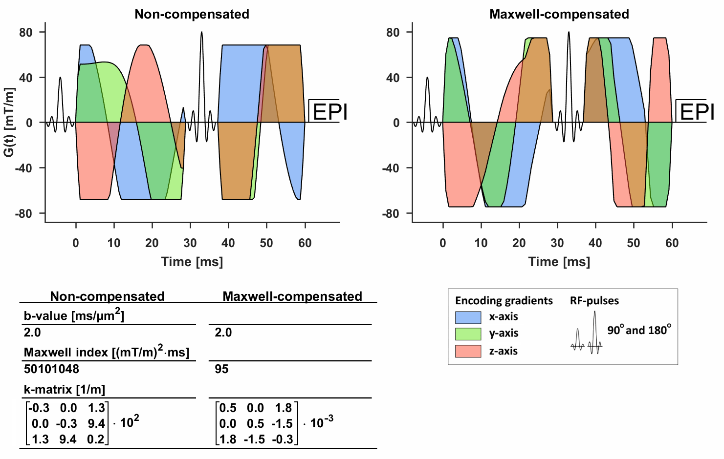
We seek a Maxwell-compensated gradient waveform $$$\mathbf{G}(t)$$$ for which ($$$\mathbf{k}=0$$$) so that $$$\text{AF}=1$$$. We propose to do this by minimizing the “Maxwell index”, defined here as the invariant scalar $$$m=(\text{Tr}[\mathbf{MM}])^{1/2}$$$, where
$$\mathbf{M}=\int_{\text{P1}}^{}\mathbf{G}(t)\mathbf{G}(t)^\text{T}\text{d}t-\int_{\text{P2}}^{}\mathbf{G}(t)\mathbf{G}(t)^\text{T}\text{d}t.\quad\text{Eq.4}$$
Importantly, $$$m$$$ is invariant to waveform rotation and when $$$m=0$$$ we know that $$$\mathbf{k}=0$$$ and $$$\text{AF}=1$$$ (unlike minimizing e.g. elements of $$$\mathbf{k}$$$). We include the minimization of $$$m$$$ in the waveform optimization framework developed by Sjölund et al.1 (https://github.com/jsjol/NOW). Note that for sequences with multiple refocusing pulses, Eq.4 is extended with additional integration periods.
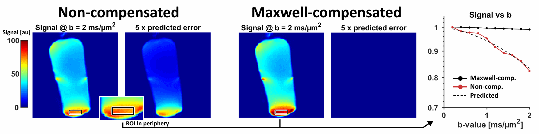
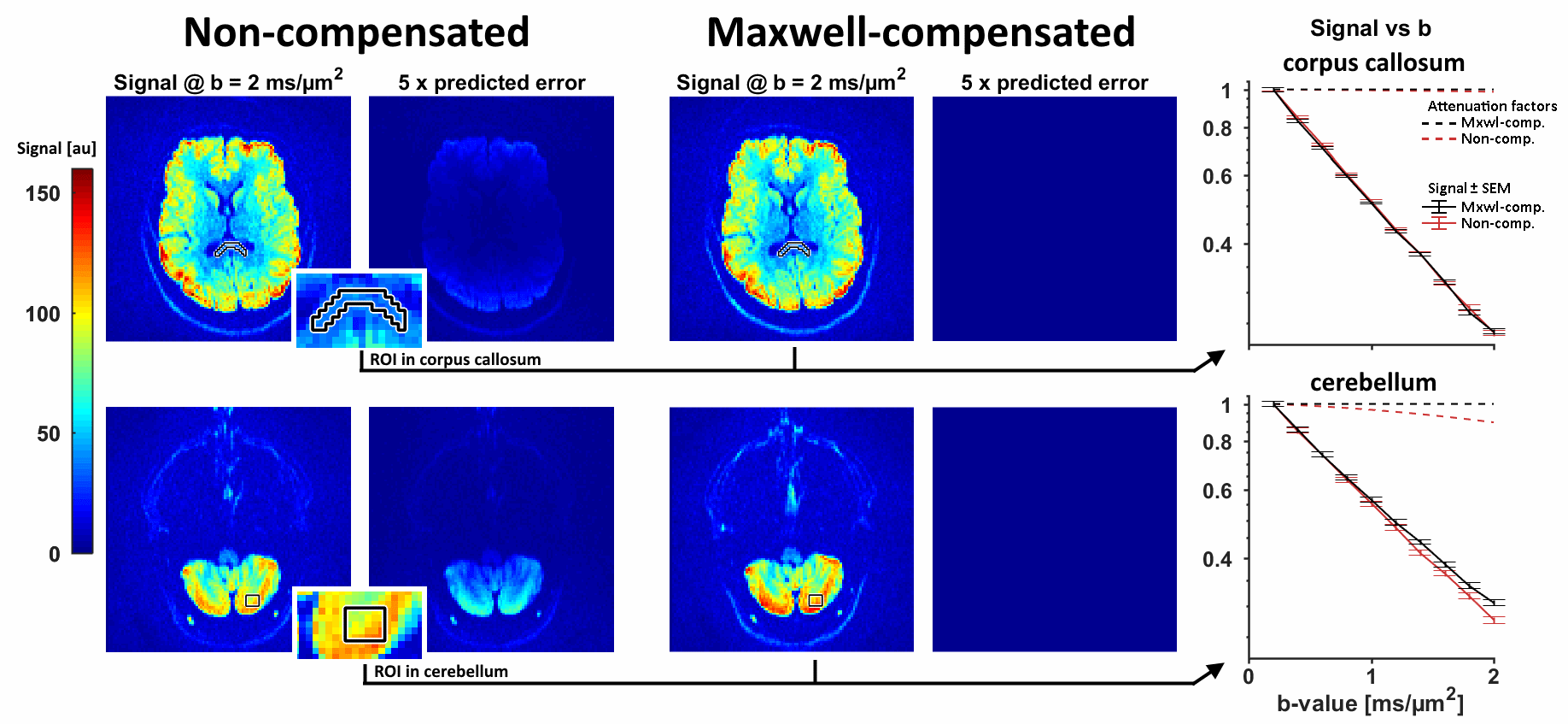
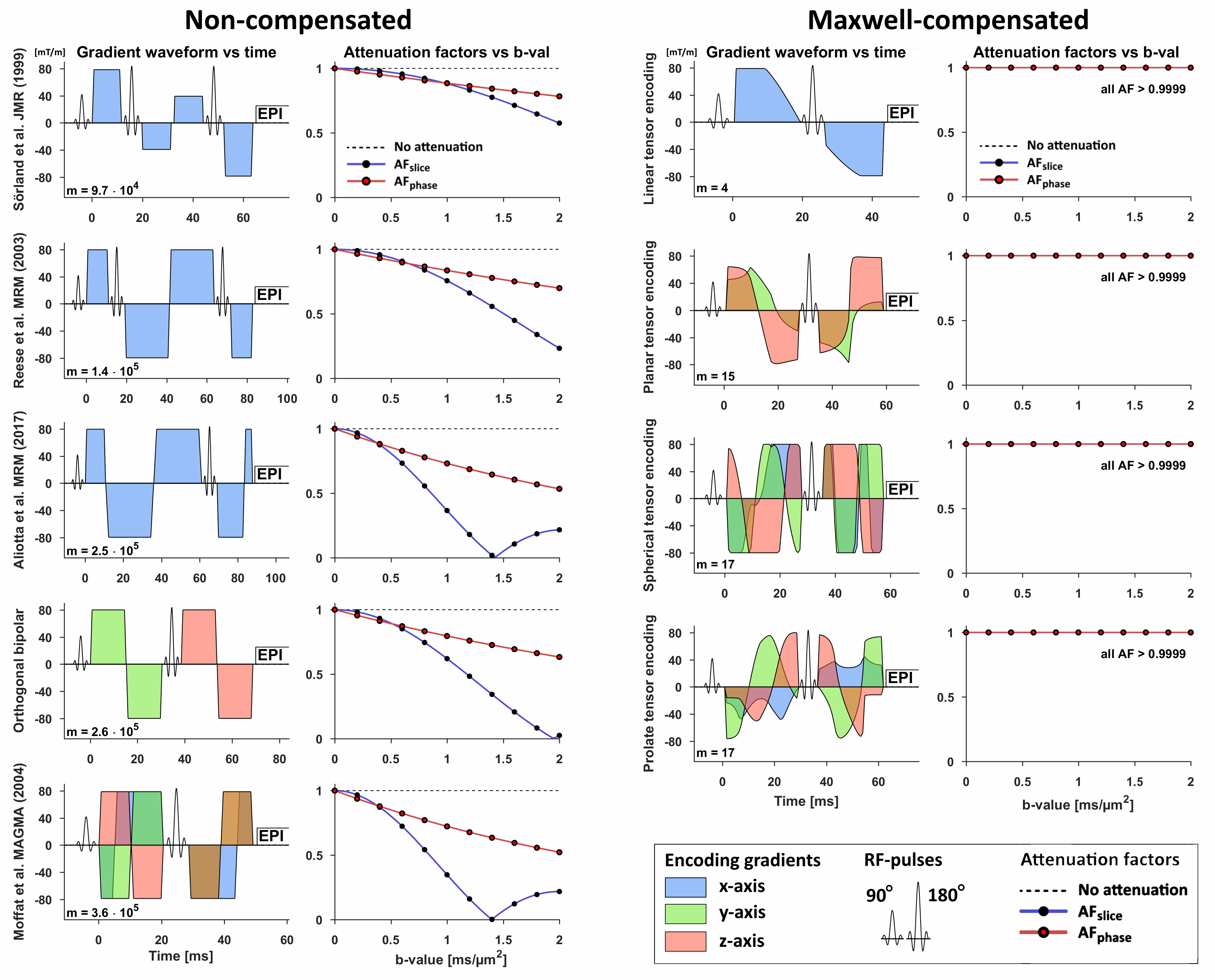
Maxwell-compensated waveforms (Fig.1) that yield spherical tensor encoding (isotropic) were validated in an oil phantom and in a healthy volunteer. Imaging was performed on a 3T MAGNETOM Prisma (Siemens Healthcare GmbH, Erlangen, Germany) with a prototype spin-echo sequence that enables arbitrary gradient waveforms; using TE=89 ms, TR=5 s, FOV=224×224 mm2, slices=30, resolution=2×2×4 mm3, iPAT=2, ∆t=0.65ms, partial-Fourier=6/8, and ten equidistant b-values between 0.2–2.0 ms/µm2. The waveforms were rotated according to Fig.1, and $$$\mathbf{n}_\text{phase}$$$ and $$$\mathbf{n}_\text{slice}$$$ were along the $$$y$$$ and $$$z$$$ axes.
Furthermore, we investigated the impact of Maxwell terms on several asymmetric waveforms found in litterature1-3,8,9. The “worst case” within 100 mm of the isocenter was estimated by independently rotating the waveform and phase/slice directions (kept orthogonal) until the minimal $$$\text{AF}$$$ was found. Analysis was performed in Matlab (The MathWorks, Natick, MA, USA).
Results
The simulations in Fig.2 show that the non-compensated design suffers gros signal error, especially at locations further away from the isocenter. By contrast, Maxwell-compensated waveforms showed negligible effects in all voxels (Fig.2). Measurements in oil (Fig.3) and in brain tissue (Fig.4) confirmed the efficacy of Maxwell-compensation, and visualize the impact of non-compensated waveforms across the FOV. For the current setup, we suggest $$$m<100$$$ (mT/m)2ms as a conservative threshold, for which $$$\text{AF}>0.999$$$ within 250 mm of the isocenter. The Maxwell-compensated waveform required approximately 3.5 ms longer echo time compared to a similar non-compensated waveform. This cost is small compared to using mirror-symmetric (+18 ms) or repeated waveform designs (+40 ms)1. Finally, Figure 5 shows that Maxwell-compensation can be achieved for arbitrary b-tensor shapes7, and that several asymmetric designs from literature may be affected by Maxwell terms.
Discussion and conclusion
Signal errors and artifacts caused by Maxwell terms are a critical consideration in diffusion encoding with asymmetric waveforms. We have showed that such effects can be mitigated by minimizing the Maxwell index, even for arbitrary waveform rotations (encoding directions) and oblique phase/slice directions. Although prospective and post-hoc corrections are possible in many scenarios4, a bias-free acquisition may be preferable, since an accurate correction requires detailed knowledge of the imaging sequence and tissue T2*. Maxwell-compensation does introduce a small penalty to the required encoding time, but it enables asymmetric waveforms with superior efficiency1.
In conclusion, we have proposed a low-cost solution to Maxwell terms in asymmetric waveform design. This approach facilitates efficient diffusion encoding, requires no further correction, and is especially relevant for diffusion imaging at high gradient strength, low main magnetic field, large/oblique/off-isocenter FOV, and/or short T2*.
Acknowledgements
We thank Siemens Healthcare for access to the pulse sequence programming environment.References
1. Sjölund et al., Constrained optimization of gradient waveforms for generalized diffusion encoding. J Magn Reson, 2015. 261: p. 157-168.
2. Reese et al., Reduction of eddy-current-induced distortion in diffusion MRI using a twice-refocused spin echo. Magn Reson Med, 2003. 49(1): p. 177-82.
3. Aliotta et al., Convex optimized diffusion encoding (CODE) gradient waveforms for minimum echo time and bulk motion-compensated diffusion-weighted MRI. Magn Reson Med, 2017. 77(2): p. 717-729.
4. Baron et al., The effect of concomitant gradient fields on diffusion tensor imaging. Magn Reson Med, 2012. 68(4): p. 1190-201.
5. Bernstein et al., Concomitant gradient terms in phase contrast MR: analysis and correction. Magn Reson Med, 1998. 39(2): p. 300-8.
6. Meier et al., Concomitant field terms for asymmetric gradient coils: consequences for diffusion, flow, and echo-planar imaging. Magn Reson Med, 2008. 60(1): p. 128-34.
7. Westin et al., Q-space trajectory imaging for multidimensional diffusion MRI of the human brain. Neuroimage, 2016. 135: p. 345-62.
8. Sörland et al., A Pulsed Field Gradient Spin-Echo Method for Diffusion Measurements in the Presence of Internal Gradients. J Magn Reson, 1999. 137(2): p. 397-401.
9. Moffat et al., Diffusion imaging for evaluation of tumor therapies in preclinical animal models. MAGMA, 2004. 17(3-6): p. 249-59.
Figures