0186
Acceleration of Vessel-Selective 4D MR Angiography by pCASL in combination with Acquisition of Control and Labeled Images in the Same Shot (ACTRESS)1C.J. Gorter Center for High Field MRI, Department of Radiology, Leiden University Medical Center, Leiden, Netherlands, 2FMRIB Centre, Nuffield Department of Clinical Neurosciences, University of Oxford, Oxford, United Kingdom
Synopsis
In the last decade, MR dynamic angiography (4D-MRA) using arterial spin labeling has become an important alternative to contrast-enhanced 4D-MRA, although scan-time is usually much longer than contrast-enhanced 4D-MRA. Among other advantages, it has the attractive possibility to allow for vessel-selective visualization. In this study, we propose an adaptation of ACTRESS (Acquisition of ConTRol and labeled Images in the Same Shot) approach for pCASL to enable vessel-selective 4D-MRA with almost halved scan-time. In an in-vivo study, it was shown that pCASL-ACTRESS approach provided vessel-selective 4D-MRA with comparable image quality to a conventional pCASL-approach, but acquired in approximately half the scan-time.
Purpose
In the last decade, MR dynamic angiography (4D-MRA) using arterial spin labeling (ASL) has become an important alternative to contrast-enhanced 4D-MRA, by demonstrating several advantages such as no need for contrast agent, higher temporal and spatial resolution1, and the ability to perform vessel-selective MRA2. In ASL-based 4D-MRA, however, scan-time is usually much longer than contrast-enhanced 4D-MRA, not only to achieve higher temporal and spatial resolution, but also because ASL requires acquisition of two image types, i.e. labeled and control images, to subtract out static tissue signal.
“Acquisition of ConTRol and labEled Images in the Same Shot” (ACTRESS) has been proposed to shorten scan-time of ASL-based 4D-MRA by nearly a factor of two, by acquiring labeled and control images within the same Look-Locker readout (instead of two separate acquisitions)3. However, the original ACTRESS-approach is only applicable with pulsed-ASL, thereby excluding the use of pCASL-based vessel-selective approaches for vessel-specific 4D-MRA, which could be useful in many cerebrovascular diseases.
In this study, we propose pCASL-ACTRESS, which allows vessel-selective 4D-MRA, while almost halving the scan-time as compared to conventional pCASL 4D-MRA.
Methods
The ACTRESS-approach is based upon the idea of performing subtraction between images with different post-label delays (PLDs) as acquired within a single Look-Locker readout4, i.e. by performing the labeling before the 2nd-phase, the 1st-phase can be used as control image. This approach alleviates the necessity to acquire complete Look-Locker series twice for label and control conditions separately. To achieve proper background signal subtraction in ACTRESS it is essential to keep the background tissue signal-intensity as flat as possible across all Look-Locker phases. With pCASL, however, a long labeling period will result in a much longer relaxation time before starting the Look-Locker readout as compared to the Look-Locker interval, thereby destroying the steady-state condition. This will cause large signal changes of static tissue across different phases, eventually resulting in elevated background signal after subtraction, which might especially impair the visibility of peripheral arteries. To minimize this effect, a WET saturation scheme was inserted before the labeling and a variable flip-angle sweep was adopted for the excitation pulses of the different phases. Finally, Figure-1 illustrates new subtraction schemes as proposed in this study to obtain the 4D-MRA images.
Bloch equation simulations were performed to optimize parameters and subsequently these settings were tested in four healthy volunteers: labeling duration of 800ms, PLD of 200ms, 10 Look-Locker phases with 180ms temporal resolution. For readout a 3D-TFEPI sequence was used with a TFE factor of 12 (including 2 start-up echoes) and EPI factor of 7, FOV of 220×220mm, 178×178 matrix (reconstructed at 256×256), 140 slices with a thickness of 0.65mm. Two excitation flip-angle schemes were tested: constant flip-angle of 8° across all phases, and a flip-angle sweep with 7° and 7.5° for the 1st and 2nd-phase (and 8° for all others). The scan-time of the conventional approach (separate label and control acquisition, as acquired for comparison) and ACTRESS-approach were 5min 19s and 2min 40s, respectively.
Results
Bloch equation simulations suggested that background tissue signal-intensity will be constant over the Look-Locker phases when using the parameters as stated above (Figure-2a). In the in-vivo study, however, magnetization transfer effects from the labeling pulses imposed an additional signal-intensity modulation (Figure-2b). By applying the flip-angle sweep, the signal-intensity changes between the 2nd- and last-phase were smaller, although a larger signal-intensity difference was observed between the 1st- and 2nd-phase, which is considered less crucial, because the early inflow-phases have the highest ASL-signal.
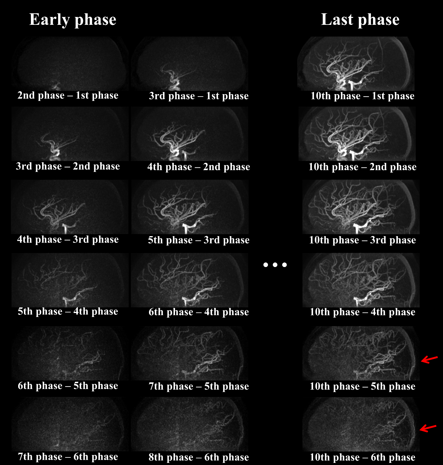
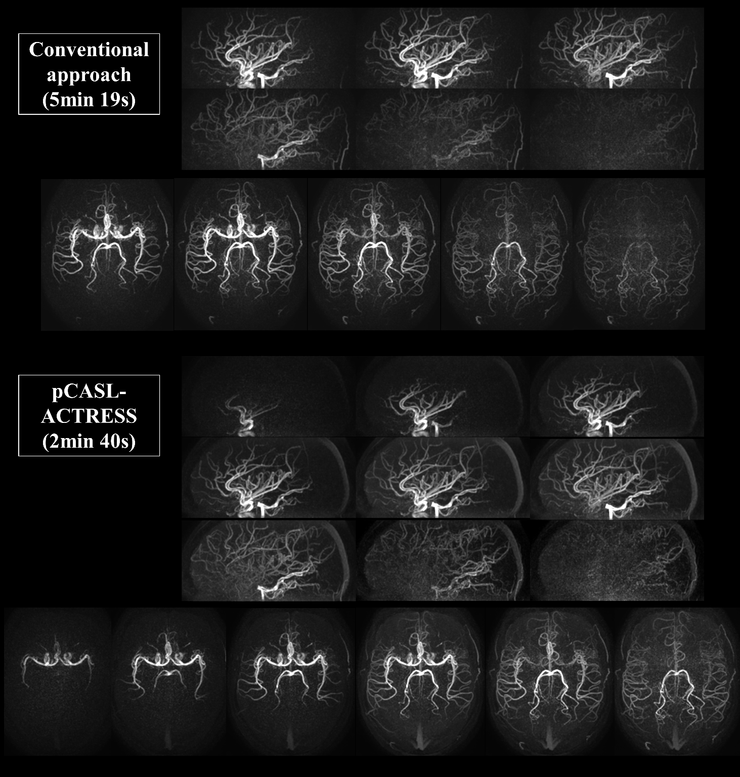
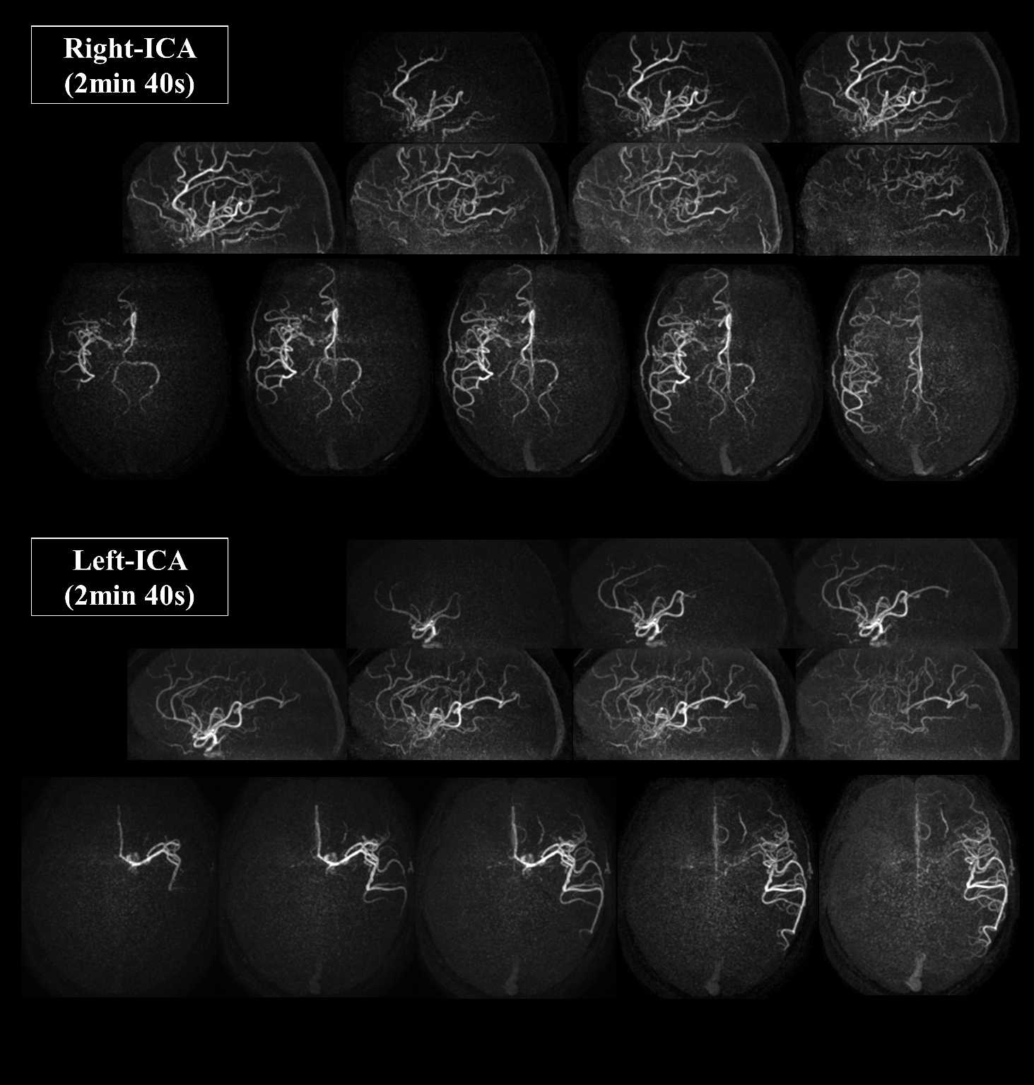
Figure-3 shows representative images of the early inflow-phase and the last peripheral-phase obtained by the proposed subtraction schemes. By using a later phase as control image for subtraction, slowly arriving arterial blood such as those in the occipital artery were better visualized (indicated by arrows). Although a slight elevation of background signal was observed in the ACTRESS-approach compared to the conventional approach (Figure-4), visualization of distal arteries was not hindered for any case. Finally, Figure-5 shows representative images of vessel-selective 4D-MRA acquired in 2min 40s.
Discussion and conclusion
Multiple subtraction schemes were proposed for two reasons: (i) slowly flowing labeled blood arriving later than 1st-phase could be visualized by using a later phase as control image, and (ii) subtraction using neighboring phases could minimize differences in background signal-intensity. This resulted indeed in better visualization of peripheral arteries.
In this study, pCASL-ACTRESS provided 4D-MRA with comparable image quality to the conventional pCASL approach, but acquired in approximately half the scan-time, whilst also allowing vessel-selective 4D-MRA. This approach improves the clinical usability for many cerebrovascular diseases, and can be an important asset for clinical applications.
Acknowledgements
This research was supported by the EU under the Horizon2020 program (project: CDS-QUAMRI).References
1. Uchino H, et al. A novel application of four-dimensional magnetic resonance angiography using an arterial spin labeling technique for noninvasive diagnosis of Moyamoya disease. Clin Neurol Neurosurg 2015;137:105-111.
2. Fujima N, et al. Utility of noncontrast-enhanced time-resolved four-dimensional MR angiography with a vessel-selective technique for intracranial arteriovenous malformations. J Magn Reson Imaging 2016;44(4):834-845.
3. Suzuki Y, et al. Acceleration of ASL-based time-resolved MR angiography by acquisition of control and labeled images in the same shot (ACTRESS). Magn Reson Med 2017.
4. Gunther M, et al. Arterial spin labeling in combination with a look-locker sampling strategy: inflow turbo-sampling EPI-FAIR (ITS-FAIR). Magn Reson Med 2001;46(5):974-984.
Figures