0185
Investigation of Intracranial Artery Selective Visualization in Superselective 4D-MR Angiography with Pseudo-Continuous Arterial Spin Labeling Combined with CENTRA-Keyhole and View-sharing (SS-4D-PACK)1Philips Japan, Tokyo, Japan, 2Department of Clinical Radiology, Graduate School of Medical Sciences, Kyushu University, Fukuoka, Japan, 3Philips Research, Hamburg, Germany, 4Division of Radiology, Department of Medical Technology, Kyushu University Hospital, Fukuoka, Japan, 5Philips Healthtech, Tokyo, Japan
Synopsis
Four dimensional (4D) MR Angiography with Pseudo-Continuous Arterial Spin Labeling (pCASL) combined with CENTRA-Keyhole and View-sharing (4D-PACK) has demonstrated high flow visualization ability in clinical use. In this study, we combined Superselective-pCASL with 4D-PACK (SS-4D-PACK) and investigated artery-selective visualization ability in SS-4D-PACK through comparison with contrast inherent inflow enhanced multi-phase angiography combining vessel-selective arterial spin labeling technique (CINEMA-Select). SS-4D-PACK showed higher vessel selectivity and vessel visualization in peripheral artery compared with CINEMA-Select.
Purpose
Four dimensional (4D) MR Angiography based on Pseudo-Continuous Arterial Spin Labeling (pCASL) combined with CENTRA-Keyhole and View-sharing (4D-PACK) has been proposed1. Here, acquisition times are accelerated by using Keyhole and View-sharing and showed high flow visualization capability1. Recently, Superselective-pCASL was introduced as a vessel-selective labeling technique2,3. In this study, we combined Superselective-pCASL with 4D-PACK (SS-4D-PACK) and compared its ability to visualize individual cerebrovascular arteries with an existing selective 4D-MRA approach named contrast inherent inflow-enhanced multi-phase angiography combining vessel-selective arterial spin labeling technique (CINEMA-Select) 4, 5.Methods
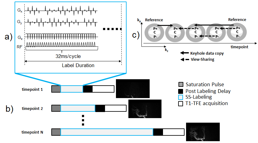
SS-4D-PACK-scheme
SS-4D-PACK scheme is visualized in Figure 1. On top of the non-selective pCASL that uses gradient Gz in the direction of the blood flow, the gradients perpendicular to the flow direction (Gx, Gy) are added in SS-4D-PACK (Figure 1a). By dynamically changing the direction of the additional gradients, a circular or elliptical labeling spot can be created. The size of the labeling focus can be adjusted by changing the zeroth moment of the transverse gradients; hence this method is able to label individual vessels. SS-pCASL labeling duration (LD) is changed dynamically to achieve 4D-MRA images and LD plus post labeling delay defines the respective timepoint (Figure 1b). Keyhole and View-sharing scheme used in SS-4D-PACK is described in Figure 1c. Two reference images at the first and the last timepoint were chosen. A central disk is delineated elliptically by the Keyhole percentage (Keyhole%) and subdivided into three regions, namely P+, C, and P–. While the central region C is acquired at every timepoint, the peripheral regions P+ and P– are acquired in an alternating fashion, and missing profiles are copied from the next timepoint data.
Magnetic Resonance (MR) Experiments
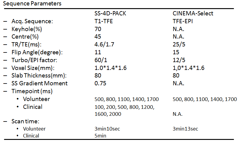
SS-4D-PACK scheme was implemented on a 3.0T Ingenia scanner (Philips, Best, The Netherlands). Left and right Internal Carotid Artery (ICA) circulations in three healthy subjects (3 Males, mean age 43.0 years, total of six ICAs labeled) were examined with SS-4D-PACK and CINEMA-Select. In addition, SS-4D-PACK was applied in a patient (female with age 69 years) underwent an extracranial-intracranial high-flow bypass using a radial graft and the proximal ligation of the left ICA for a giant aneurysm. The labeling focus was placed on the proximal left external carotid artery (ECA) for this patient. Sequence parameters used in this study are shown in Figure 2. Informed consent required by the Institutional Review Board was obtained. For image evaluation, 80mm slab maximum intensity projection images were used.
Validation of flow visualization
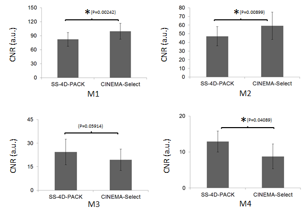
For the assessment of flow visualization ability, circular regions of interest (ROI) were selected from the white matter (WM) and the middle cerebral artery (MCA). Four MCA ROIs were placed on M1, M2, M3, and M4 branches. The contrast-to-noise ratio (CNR) between MCA and WM was measured using the following equation: 6
CNR = (bloodmax–WMave)/WMSD.
Here, bloodmax is the maximum signal in all ROIs over the acquired timepoints. WMave and WMSD are the average signal and standard deviation, respectively, in the WM ROI. The paired T-test was used in the statistical analysis and p<0.05 was considered statistically significant.
Results and Discussion
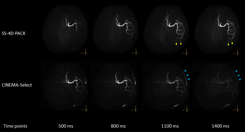
The representative 4D-MRA in right ICA circulation generated from SS-4D-PACK and CINEMA-Select are shown in Figure 3. The images obtained by SS-4D-PACK exhibit high flow visualization in peripheral areas (yellow arrows). In addition, ECA circulation visualized in CINEMA-Select (blue arrows) was not observed in SS-4D-PACK, which indicates high vessel selectivity.
Assessment of Flow Visualization Ability
The average CNRs in each MCA segment are shown in Figure 4. The CNRs in CINEMA-Select were significantly higher in M1 and M2 branch compared with SS-4D-PACK. On the other hand, CNRs in SS-4D-PACK tend to be higher (P=0.05914) compared with CINEMA-Select in M3 and there was significant difference in CNR in M4. Sufficient signal from labeled blood at late timepoints is important for the visualization of small intracranial arteries or collateral flow. Moreover, disease-related slow hemodynamics may be challenging to visualize over the whole vasculature, however, the application of SS-4D-PACK in such cases seems promising.
Demonstration in Patients
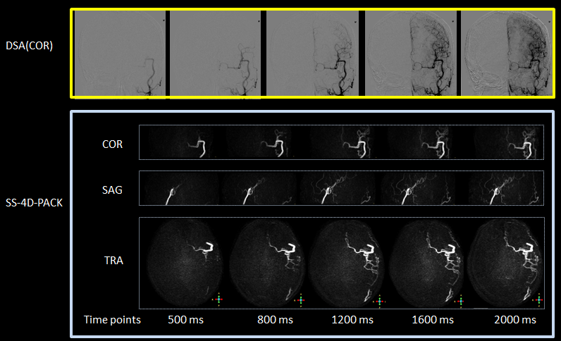
Figure 5 shows SS-4D-PACK images in a patient. SS-4D-PACK selectively visualizes the bypass graft and blood flow into the left MCA and anterior cerebral artery (ACA) territories. Visualizations of vessels other than interest were not recognized.
Conclusion
In this study, a new approach based on SS-4D-PACK has been demonstrated. SS-4D-PACK showed higher vessel selectivity and vessel visualization in peripheral arteries compared with CINEMA-Select. Further clinical investigation with larger clinical cases is mandatory.Acknowledgements
No acknowledgement found.References
- Osamu Togao, Akio Hiwatashi, Makoto Obara, et al. 4D MR Angiography with Pseudo-Continuous Arterial Spin Labelling Combined with CENTRA-Keyhole (4D-PACK): Visualization of Distal Cerebral Arteries in Moyamoya Disease. Proceedings of the 25th Annual Meeting of ISMRM, Honolulu, 2017 #145
- Michael Helle, David G. Norris, Susanne Rufer, et al. Superselective Pseudocontinuous Arterial Spin Labeling. Magn Reson Med 2010;64: 777–786.
- Ulf Jensen-Kondering, Thomas Lindner, Matthias J.P. van Osch, et al. Superselective pseudo-continuous arterial spin labeling angiography. European Journal of Radiology 2015;84: 1758–1767.
- Masanobu Nakamura, Masami Yoneyama, Takashi Tabuchi, et al. Vessel-selective, non-contrast enhanced, time-resolved MR angiography with vessel-selective arterial spin labeling technique (CINEMA-SELECT) in intracranial arteries. Radiol Phys Technol. 2013;6: 327-334.
- Noriyuki Fujima, Toshiya Osanai, Yukie Shimizu, et al. Utility of noncontrast-enhanced time-resolved four-dimensional MR angiography with a vessel-selective technique for intracranial arteriovenous malformations. J Magn Reson Imaging. 2016;44:834-845.
- Makoto Obara, Osamu Togao, Masami Yoneyama, et al. Acceleration-selective arterial spin labeling for intracranial MR angiography with improved visualization of cortical arteries and suppression of cortical veins. Magn Reson Med. 2017;77:1996-2004.
Figures