0146
31-channel receive coil array combined with an 8-channel whole-brain dipole transmit array1LIFMET, EPFL, Lausanne, Switzerland, 2Department of Radiology, University of Lausanne, Lausanne, Switzerland, 3Department of Radiology, University of Geneva, Geneva, Switzerland, 4CIBM-AIT, EPFL, Lausanne, Switzerland
Synopsis
To increase the parallel imaging performances while keeping high transmit field, the combination of a high-density receive coil array and a tight-fitted whole-brain dipole coil array was investigated. Measured noise correlation matrix, signal-to-noise ratio and g-factor maps were evaluated for the 31-channel receive coil array, and MRI acquisition time could be decreased up to 3.4 times without attenuation in data quality. MR images demonstrated a large spatial coverage, including cerebellum and cerebral cortex, thanks to the whole-brain dipole transmit array while the 31-channel receive coil array provided highly accelerated image acquisition.
Introduction
Receive coil arrays are known to provide high signal-to-noise ratio[1] and to enable a decreased acquisition time through parallel imaging techniques[2,3]. However, while receive coil arrays are commonly used, for head and body imaging, in combination with volume coils and transmit loop or microstrips coil arrays[4,5,6], the feasibility of using a large number of receivers with a transmit dipole coil array on the human brain at 7T is still not investigated. In this study we evaluated a 31-channel receive array with 8-channel tight-fitted transmit dipole array to acquire highly accelerated whole-brain images, including cerebellum and cerebral cortex, with high transmit efficiency at 7T.Methods
Coil Design: The transmit coil array consisted in seven dipoles and two quadrature frontal loops placed around the head in a way to achieve whole-brain coverage (Fig.1A). RF safety was evaluated with finite-difference time-domain simulations (Sim4Life 3.4,ZMT,Switzerland). A tight-fitting helmet (222mm in anterior-posterior (AP) direction,180mm left-right,231mm head-foot) was 3D-printed (EOSINT P395,EOS,Germany) in nylon (EOS,PA2200) to accommodate the head shape and to place the receivers. The dipoles were placed at a maximal distance of 15mm from the helmet and no detuning of the dipoles was implemented in MR measurements during reception. A total of 29 circular (with diameters from 70mm to 85mm) receive loop coils were built and arranged on the helmet such as their center was aligned with the dipoles, when feasible, to minimize interactions (Fig.1A-B). In addition, a PIN-diode was placed across the matching capacitor CM and connected in series with a hand-wounded inductor to enable active detuning of the receivers during RF transmission (Fig.1C). Geometric overlap was adopted to cancel the inductive coupling between neighbors. For next-nearest neighbor decoupling, preamplifier decoupling method[7] was implemented by connecting, through a half-wavelength coaxial cable, a low-input impedance preamplifier (WMM7RP,WantCom Inc.,Minnesota,USA) across the PIN-diode used for detuning to create a virtual open circuit (Fig.1B). To minimize common-modes, shielded cable traps were added on each receiver line, between the loop coil and preamplifier. The two frontal loops were used in both transmit and receive, controlled by an in-house built transmit-receive switch with quadrature hybrid.
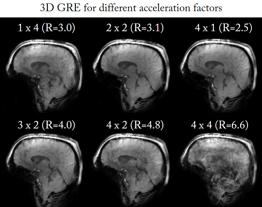
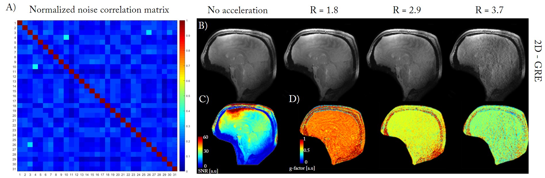
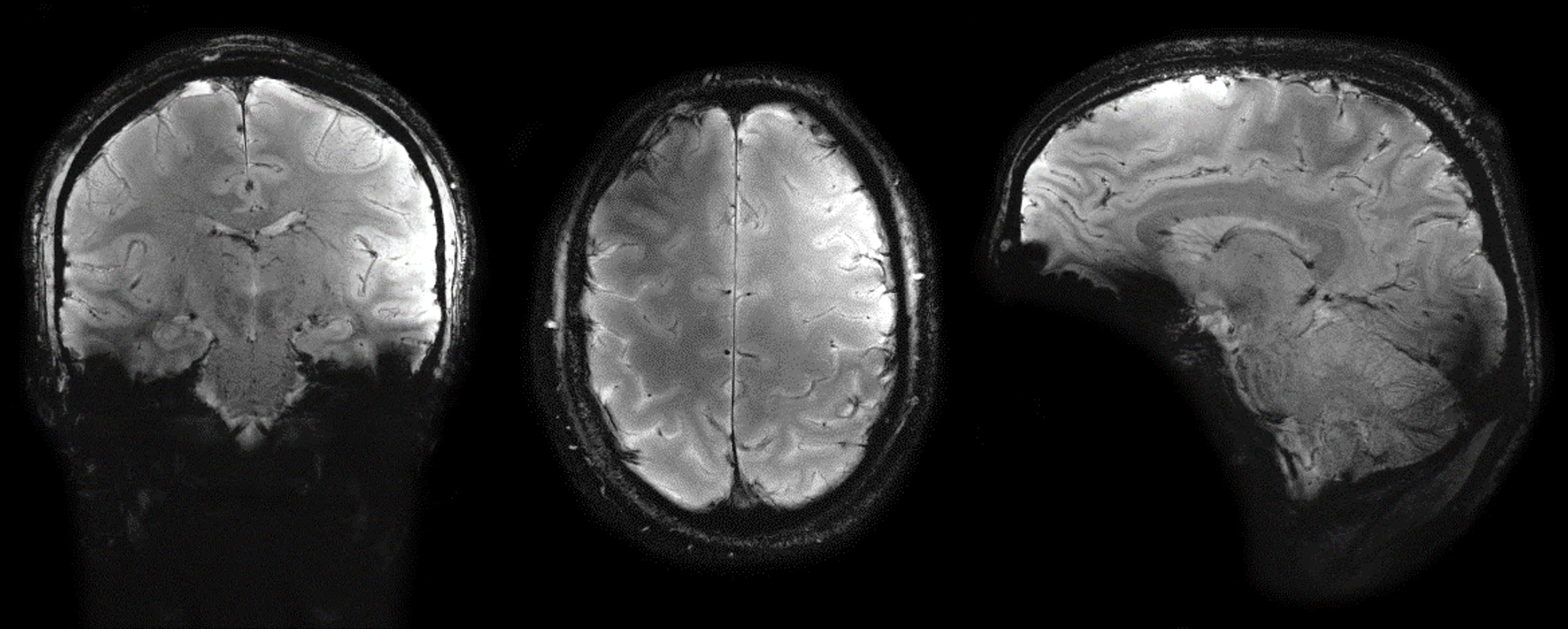
MR data was acquired from healthy volunteers using a Magnetom 7T head-only MR scanner with an 8x1kW RF-amplifier (Step 2.3,Siemens,Erlangen,Germany). Optimal transmit RF-phases were calculated with a particle-swarm optimization[8] method and applied for all measurements. 3D-GRE images (1.5x1.5x3.0mm3,TR/TE=60/4ms,R=1/3.0/3.1/2.5/4/4.8/6.6) were acquired to determine the capability for the receive array to accelerate the acquisition in multiple directions. SNR, g-factor maps and noise correlation matrix[3] (normalized to 1 for diagonal terms) were measured from a sagittal 2D-GRE image (1.0x1.0x1.0mm3,TR/TE=1000/3.37ms,FA=48°,192x192 matrix), acquired without acceleration (signal/noise) and at different acceleration factors (R=1.8/2.9/3.7,A-P,only signal). Anatomical images were obtained from a high-resolution multi-slice gradient-echo sequence at R=3.4 (0.3x0.3x3.0mm3,TR/TE=1000/16ms,TA=3min,640x640 matrix).
Results
No acceleration-related artifact was observed in 3D-GRE images for R-values up to 4 except for R=3.0 which demonstrated a blurring effect in the center of the brain (Fig.2). Noise correlation matrix showed good decoupling between all the receivers in the array (Fig.3A). Relatively low SNR values were measured with no acceleration (Fig.3C), but no variations in SNR was observed for increased R (from 1.8 to 3.7). For R= 3.7, the 2D-GRE image was visibly altered by the acceleration-related artifacts (Fig.3B). Mean g-factors over the brain were 0.75, 0.59 and 0.53 for R= 1.8, 2.9, 3.7, respectively (Fig.3D). Anatomical images showed a large coverage of the whole-brain with a good homogeneity (Fig.4) except at the bottom part of the occipital lobe where the signal appeared to be canceled.Discussion and conclusion
The feasibility of combining a high-density
receive coil array with a tight-fitted transmit dipole coil array was
demonstrated. The capability to decrease the 3D acquisition time up to almost 4
times will benefit to fMRI studies where timing of the acquisition is critical.
However, g-factor and SNR maps appeared to be relatively low, and further
optimization of the receive array still needs to be performed. Moreover, due to
the geometrical arrangement of receive loops configuration, the transmit field
was altered, which is resulted in signal voids visible at the bottom of
occipital lobe. This may originate from the shielded cable traps used on the
receive coaxials, from a non-perfect alignment of the loops below the dipoles
or from the active detuning trap. Nevertheless, high-resolution multi-slice gradient-echo
images could be acquired at R= 3.4 without visible attenuation of the data
quality. We conclude that the realization of a 31-channel receive array with an
8-channel dipole transmit array is feasible and promises highly accelerated and
transmit-efficient MR acquisitions overall the human brain at 7T.Acknowledgements
This study was supported by Centre d’Imagerie Biomedicale (CIBM) of the UNIL, UNIGE, HUG, CHUV, EPFL and the Leenaards and Jeantet Foundations.References
[1] Wiggins G.C. et al. Magn. Reson. Med. 2006; 56: 216-223
[2] Griswold M.A. et al. Magn. Reson. Med. 2002 ; 47(6) : 1202-1210
[3] Pruessmann K. et al. Magn. Reson. Med. 1999; 42: 952-962
[4] Adriany G. et al. Magn. Reson. Med. 2010; 63: 1478-1485
[5] Raghuraman S. et al. J. Magn. Reson. 2013; 238-244
[6] Zhao W. et al. Magn. Reson. Med. 2014; 72(1): 291-300
[7] Roemer PB. et al. Magn. Reson. Med. 1990
[8] Clement J. et al. Proc. ESMRMB, 2015
Figures