0141
Evaluation of Parallel Imaging performance gains with 64 channel receivers at 7 Tesla1Center for Magnetic Resonance Research, University of Minnesota, Minneapolis, MN, United States, 2Lifeservices LLC, Minneapolis, MN, United States
Synopsis
Evaluation of a 64 channel receiver relative to a 32 channel receiver shows that gains in parallel imaging performance for SMS/MB of 40 to 60% is feasible, such that, highly desirable, single-shot, multislice, whole brain coverage with <1s TR and 1mm or better isotropic resolutions would be achievable at 7T.
Purpose
At ultra high fields (UHF), the increase in SNR and the stronger coupling between RF coil and tissue allow for efficient use of smaller coils and higher coil densities. In addition, parallel imaging performance improves at UHF due to the increased spatial complexity of B1 phase and amplitude arising from the traveling wave behavior of high frequency RF in the human body(1,2). As such, synergistic combination of high number of receive channels(3,4) and UHF should be particularly beneficial for parallel imaging and image acceleration. To exploit this potential, we have recently built the first 64 channel 7Tesla receiver system (1). Here, we report on parallel imaging performance gains achievable with a novel, close fitting 64 channel fMRI coil for whole brain coverage compared to the current standard of 32 channel arrays at 7T.Methods
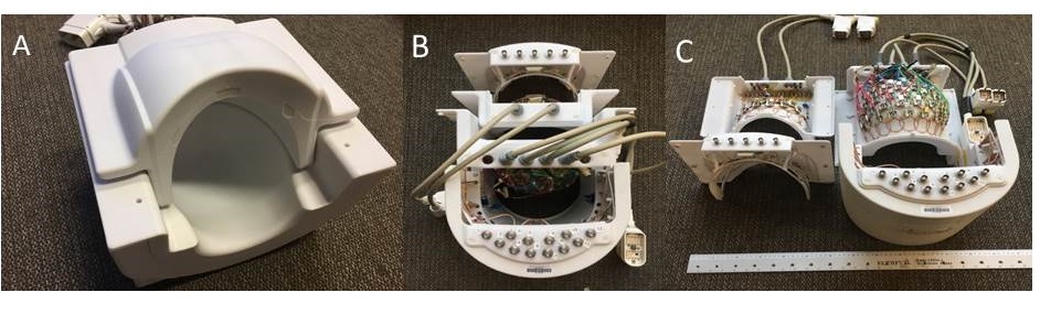
System Data was acquired on a 7T system, with a Siemens Console modified to accommodate 64 receivers (5). We compared a prototype 16-channel transmit/64-channel receive (16Tx/64Rx) coil developed jointly with Lifeservices LLC, Minneapolis MN vs. the standard product 1Tx/32Rx coil (Nova Medical, Wilmington, MA, USA). The 64channel coil is an extension of a previously presented 16Tx/32Rx open faced design (6) with similar overlapped dual row 16 channel loop transmitters (Figure 1) The coil consist of a close fitting bottom former with 48 receiver loops of 4.5cm to 5.5cm diameter size and a top former containing 16 receiver loops ranging in size from 5cm to 6cm. A total of 16 overlapped loop transmitters (~10cmx12cm) in a dual row configuration supported B1+ uniformity and SAR optimization. Acquisition was performed in accordance with local IRB oversight on 3 volunteers. For the 16Tx/64Rx coil, B1+ optimization was performed using slice specific single spoke pulses for 2D acquisitions(7). For 3D volumetric whole brain data, imaging with 2 different B1+ phase solutions was performed and combined in a phase-sensitivity approach, similar to TIAMO(8). For the 1Tx/32Rx coil, matched 3D and 2D acquisitions were performed.
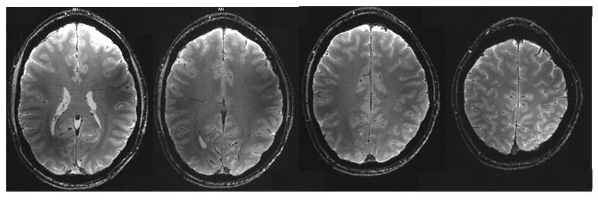
Data A fully sampled 1mm isotropic 3D GRE acquisition with TE/TR of 3ms/6.7ms and with a FOV of 256x176x256mm were obtained. A fully sampled 1mm isotropic 2D GRE with an axial/coronal oblique FOV aligned approximately with the AC-PC line, with TE/TR of 15ms/2450ms were acquired (figure 2), as well as 2D-EPI images using a multiband (MB) factor of 4, a phase-encoding undersampling factor of 3, oblique FOV, 0.8mm isotropic resolution, AP phase-encoding and LR readout. The FOV from the 3D data was tilted to an oblique FOV, and reduced to a [220x176 x140mm]
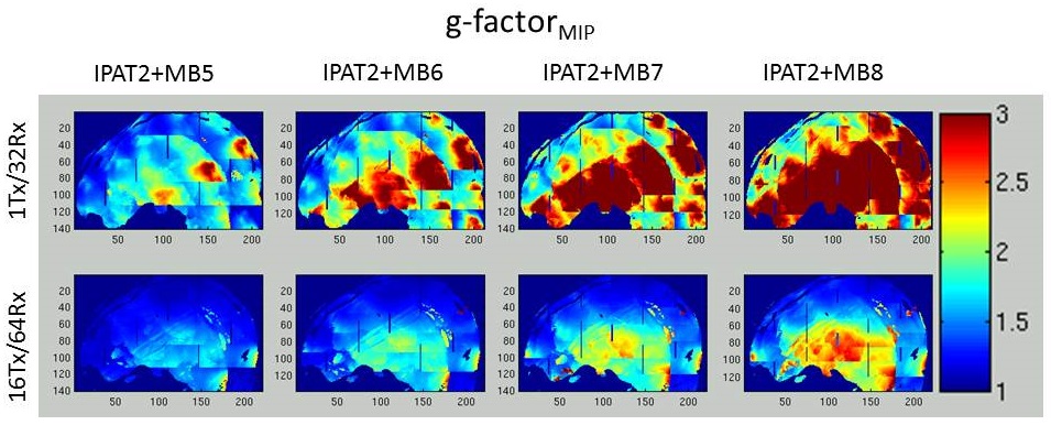
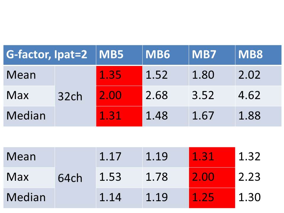
G-factors: The g-factors(9) were calculated for the 3D volume; g-factor histograms were determined, and the mean, median and 98 percentile maximum are reported. For g-factor maps, a g-factorMIP was extracted as a Maximum intensity projection in the LR direction, over a 80mm slab in the sagittal plane and displayed over a silhouette of the central sagittal slice.
Results
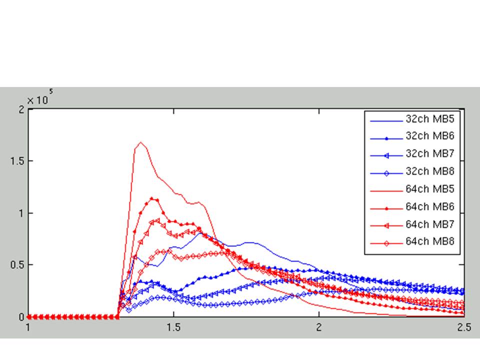
Figure 3 shows g-factorMIP for a PE undersampling of 2 (iPAT2), and MB factors of 5,6,7 and 8 (left to right) with FOV-shift 1/3, using a 1Tx/32Rx (top row) and a 16Tx/64Rx coil (bottom row), for oblique FOV slices. For iPAT2xMB5 acceleration with the 32Rx Nova coil (as employed in the Human Connectome Project (HCP) 7T resting-state fMRI (rfMRI) component), there are local hot spots of increased noise-amplification; these are removed with the 64Rx prototype coil for the same degree of acceleration. The mean, median and max g-factors are listed for the 3D whole brain g-factors maps in table 1 (figure 4), and the histograms for these maps are presented in figure 5. The mean values are similar to the median values for the 64Rx coil; however, for the 32Rx coil, the mean is higher than the median, reflecting a distribution which has a significant tail. For the 64Rx, a 40 to 60% increase in total acceleration to iPAT2xMB7 or iPAT2xMB8 can be achieved when approximately matched for the g-factor performance of the 32Rx coil at a iPAT2xMB5 performance (table1).Discussion
The 64Rx receiver array supported a 40 to 60% increase in acceleration with SMS/MB imaging compared with a 32Rx receiver array. This is a significant gain for applications that require simultaneously high temporal and spatial resolution, such as fMRI and diffusion imaging (dMRI) as employed in the HCP. With such gains, highly desirable, single-shot, multislice, whole brain coverage with <1s TR and 1mm or better isotropic resolutions would be achievable, and would substantially surpass the current HCP achievement of 1s TR and 1.6mm isotropic resolution for rfMRI at 7T. These gains would also translate into important acceleration improvements in other imaging acquisitions, especially where 2D acceleration can be employed.Acknowledgements
This work was supported by NIH U01 EB025144, S10 RR026783, BTRC P41 EB015894, P30 NS076408 and WM Keck FoundationReferences
1. Wiesinger, F., Van De Moortele, P. F., Adriany, G., De Zanche, N., Ugurbil, K. & Pruessmann, K. P. Parallel imaging performance as a function of field strength - An experimental investigation using electrodynamic scaling. (2004) Magnetic Resonance in Medicine 52, 953-964.
2. Wiesinger, F., Van de Moortele, P. F., Adriany, G., De Zanche, N., Ugurbil, K. & Pruessmann, K. P. Potential and feasibility of parallel MRI at high field. (2006) NMR in Biomedicine 19, 368-378.
3. Wiggins, G. C., Polimeni, J. R., Potthast, A., Schmitt, M., Alagappan, V. & Wald, L. L. 96-Channel receive-only head coil for 3 Tesla: design optimization and evaluation. (2009) Magn Reson Med 62, 754-762.
4. Keil, B., Blau, J. N., Biber, S., Hoecht, P., Tountcheva, V., Setsompop, K., Triantafyllou, C. & Wald, L. L. A 64-channel 3T array coil for accelerated brain MRI. (2013) Magn Reson Med 70, 248-258.
5. Auerbach E, DelaBarre L, Van de Moortele PF, Strupp J, Gumbrecht R. , Potthast A., Pirkl G, Moeller S, Hanna B, Grant A, Adriany G. Ugurbil K, An Integrated 32-Channel Transmit and 64-Channel Receive 7 Tesla MRI System, 25th Scientific Meeting of the International Society for Magnetic Resonance in Medicine (ISMRM). Honolulu (HI), 2017, p. 1218
6. Adriany, G., Schillak, S., Waks, M., Tramm, B., Grant, A., Yacoub, E., Vaughan, J. T., Olman, C., Schmitter, S. & Ugurbil K. in23rdScientific Meeting of the International Society for Magnetic Resonance in Medicine (ISMRM), Toronto, 2015, p.622
7. Wu X, Schmitter S, Auerbach EJ, Ugurbil K, Van de Moortele PF. A generalized slab-wise framework for parallel transmit multiband RF pulse design. Magn Reson Med 2016;75(4):1444-1456.
8. Orzada S, Maderwald S, Poser BA, Bitz AK, Quick HH, Ladd ME. RF excitation using time interleaved acquisition of modes (TIAMO) to address B1 inhomogeneity in high-field MRI. Magn Reson Med 2010;64(2):327-333.
9. Pruessmann KP, Weiger M, Scheidegger MB, Boesiger P. SENSE: sensitivity encoding for fast MRI. Magn Reson Med 1999;42(5):952-962.
Figures