0125
Late gadolinium enhancement of colorectal liver metastases post-chemotherapy is associated with tumour fibrosis and overall survival post-hepatectomy1Department of Medical Imaging, Sunnybrook Health Sciences Centre, University of Toronto, Toronto, ON, Canada, 2Department of Surgery, Sunnybrook Health Sciences Centre, University of Toronto, Toronto, ON, Canada, 3Department of Pathology, Sunnybrook Health Sciences Centre, University of Toronto, Toronto, ON, Canada
Synopsis
Preoperative MRI is routinely used for diagnosis, staging, and operative planning of colorectal liver metastases (CRCLM), but still relatively unexplored for preoperative prognosis. Tumour fibrosis in post-hepatectomy CRCLM specimens is associated with long-term survival and late gadolinium enhancement is associated with tumour fibrosis in other disease processes (eg. cholangiocarcinoma). We performed a retrospective cohort study (n=121) in patients who received a clinical gadolinium-enhanced MRI prior to hepatectomy for CRCLM. We determined that strong enhancement on delayed phase MRI was associated with tumour fibrosis post-hepatectomy and overall survival.
Introduction
Colorectal cancer is the second leading cause of cancer deaths in the developed world (1). Approximately half of patients develop liver metastases and most deaths are related to metastatic disease (2). The ability to predict prognosis informs treatment recommendations, including surgery and/or chemotherapy. MRI is routinely used clinically for diagnosis, staging, and operative planning in patients being considered for liver resection, but the use of MRI as a prognostic biomarker in this setting is relatively unexplored. Tumour fibrosis in post-heptectomy colorectal liver metastases (CRCLM) specimens is associated with overall survival (3-4). Late gadolinium enhancement is associated with tumour fibrosis in other diseases such as cholangiocarcinoma (5).Materials and Methods
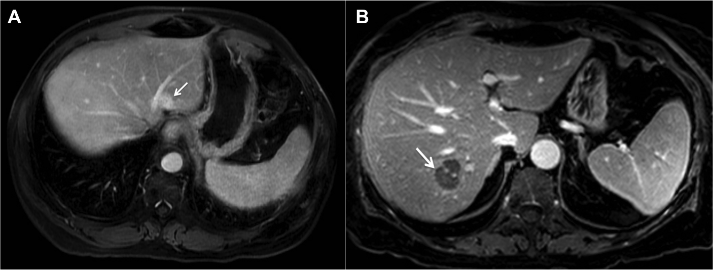
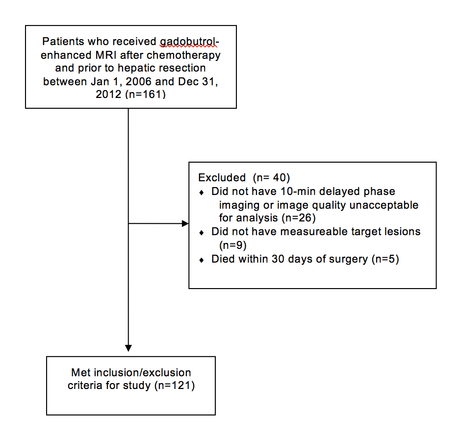
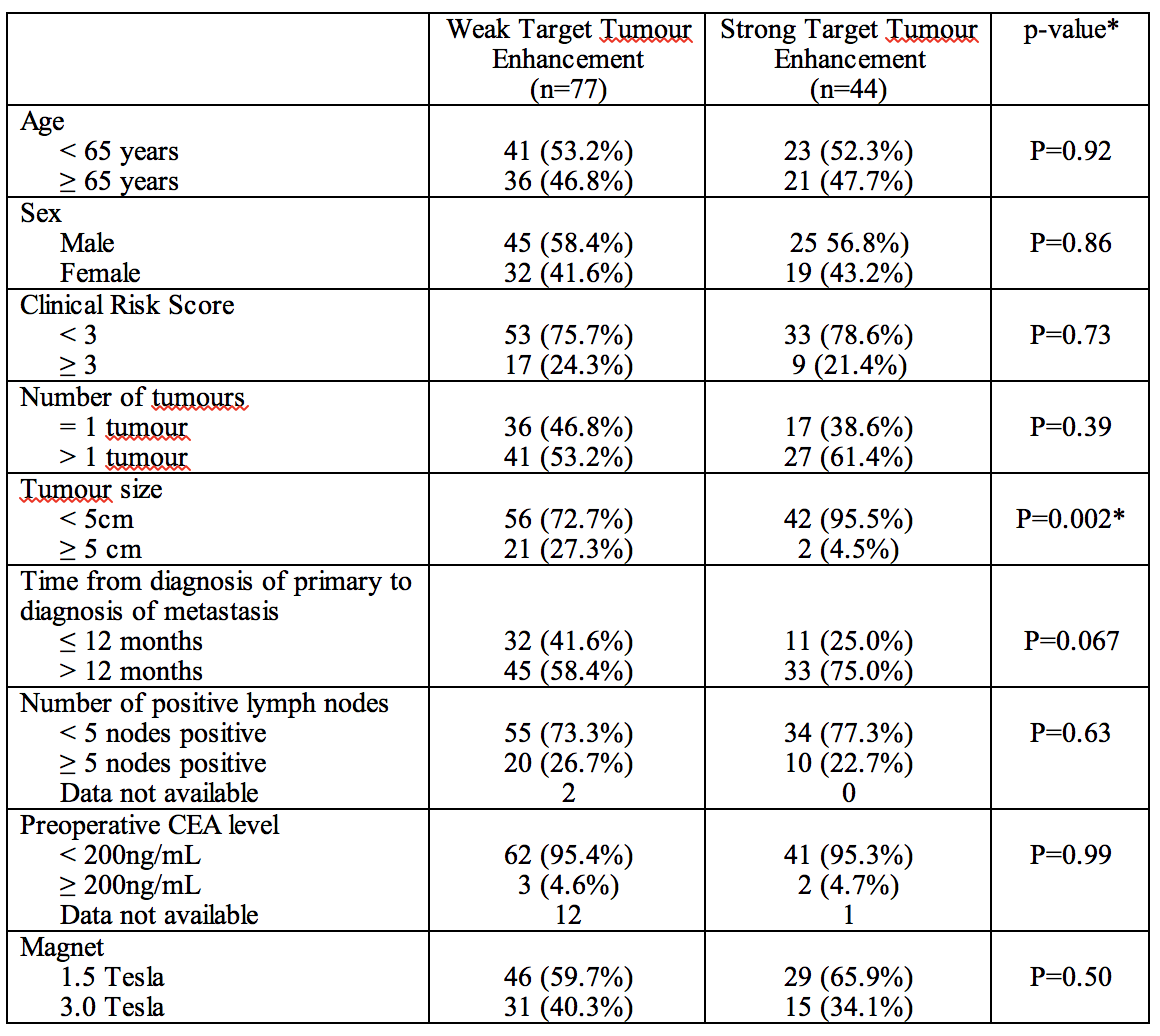
The institutional review board approved this retrospective cohort study and waived the requirement for informed consent. A cohort of 121 surgical patients who received preoperative MRI after chemotherapy between 2006-2012 were included in this study. Target tumour enhancement (TTE), defined as the mean contrast-to-noise ratio of up to 2 target lesions on late-phase gadobutrol-enhanced MRI, was determined by two independent raters. The average TTE was correlated with tumour fibrosis on post-hepatectomy specimens using Spearman correlation and with survival post-hepatectomy using Kaplan-Meier and Cox-Regression. Inter-rater reliability was determined using relative intra-class correlation coefficients.Results
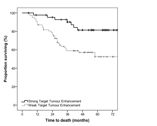
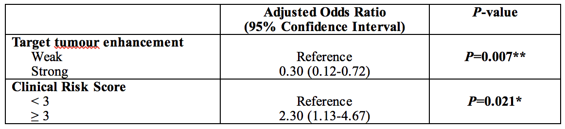
In the surgical cohort (mean age: 63.0 years; male: 70), TTE was associated with tumour fibrosis (R=0.44, p<0.001). Strong TTE was associated with improved survival compared to weak TTE (3-year survival: 90.1% vs. 58.9%, p=0.003) with a hazard ratio of 0.30 (95% CI: 0.12-0.72, p=0.007), after adjusting for known prognostic variables (6). Inter-rater reliability was very good with a relative intraclass correlation of 0.84 (95% CI: 0.77-0.89).Conclusion
Late gadolinium MRI enhancement of CRCLM post-chemotherapy is associated with tumour fibrosis and survival.Study Aim
The aim of this study was to determine whether late gadolinium MRI enhancement of colorectal liver metastases (CRCLM) post-chemotherapy is associated with tumour fibrosis and survival post-hepatectomy.Acknowledgements
Thank you to Dr. Pascal Tyrell for statistical support and to Mr. Thayalasuthan Vivekanandan for administrative support.References
(1) Torre LA, Bray F, Siegel RL, Ferlay J, Lortet-Tieulent J, Jemal A (2015). Global cancer statistics, 2012. CA Cancer J Clin 65:87-108
(2) Kanas GP, Taylor A, Primrose JN, et al (2012). Survival after liver resection in metastatic colorectal cancer: review and meta-analysis of prognostic factors. Clin Epidemiol 4:283-301.
(3) Poultsides GA, Bao F, Servais EL, et al (2012). Pathologic response to preoperative chemotherapy in colorectal liver metastases: fibrosis, not necrosis, predicts outcome. Ann Surg Oncol 19:2797-804.
(4) Rubbia-Brandt L, Giostra E, Brezault C, et al (2007). Importance of histological tumor response assessment in predicting the outcome in patients with colorectal liver metastases treated with neo-adjuvant chemotherapy followed by liver surgery. Ann Oncol 18:299-304.
(5) Maetani Y, Itoh K, Watanabe C, et al (2001). MR imaging of intrahepatic cholangiocarcinoma with pathologic correlation (2001). AJR Am J Roentgenol 176:1499-507.
(6) Feroci F, Fong Y (2010). Use of clinical score to stage and predict outcome of hepatic resection of metastatic colorectal cancer. J Surg Oncol 102:914-21.
Figures