0104
Hyperpolarized Xe-129 Imaging of Pluripotent Stem Cell-Derived Alveolar-Like Macrophages in the Lungs: Proof-of-Concept Study Using Superparamagnetic Iron-Oxide NanoparticlesVlora Riberdy1,2, Michael Litvack2, Elaine Stirrat2, Marcus Couch2, Martin Post2, and Giles Santyr1,2
1Department of Medical Biophysics, University of Toronto, Toronto, ON, Canada, 2Translational Medicine, The Hospital for Sick Children, Toronto, ON, Canada
Synopsis
A promising approach to treatment of chronic lung diseases is the intratracheal delivery of pluripotent stem cell derived alveolar-like macrophages (PSC-ALMs) into the injured lung to facilitate repair of damaged tissue. This treatment is hindered by the inability to assess where in the lung these cells end up. Here, hyperpolarized Xe-129 MRI paired with iron-labeled cells was used to demonstrate a proof-of-concept visualization of cells introduced in the lungs of rats. Signal hypointensities were observed at least one hour after instillation of approximately two million labeled cells in one rat, compared to instillation of control solutions in separate rats.
Introduction
Stem cells are a potentially useful treatment for chronic lung diseases, such as asthma, chronic obstructive pulmonary disease (COPD) and bronchopulmonary dysplasia (BPD).1 It has been shown that the major innate immune cells in the lungs, alveolar macrophages, can be derived from pluripotent embryonic stem cells and these alveolar-like macrophages (ALMs) promote repair of lung disease in animal models.2 Translation of this approach to the clinic will benefit from imaging methods that can detect and monitor ALMs in vivo following instillation in the lungs. It has been demonstrated that superparamagnetic iron oxide nanoparticles (SPIONs) can enable proton MRI of cells in the lung.3 Hyperpolarized (HP) MRI provides a further improvement in detection sensitivity of SPION-labeled cells in the lung.4 In this work, we demonstrate the use of HP 129Xe MRI combined with SPION labeling for detection of ALMs in the rat lung.Methods
ALMs were produced following the method of Litvack et al2 and loaded with varying concentrations of green fluorescent-labeled SPIONs (Molday ION EverGreen™). After four hours, confocal fluorescence microscopy and flow cytometry were used to confirm SPION uptake by the cells. The effects of SPIONs on cell viability after incubation with 0, 0.5, 1, 2, and 4% SPIONs (v/v) for four hours was measured using a Presto Blue® assay. As a preliminary step in the detection of ALMs in the lung, the effects of localized instillation of (i) phosphate buffer saline (PBS), (ii) PBS + SPIONs, and (iii) SPION-labeled ALMs on HP 129Xe signal acquired from the lungs of separate healthy Sprague Dawley rats in vivo were investigated. ALMs were incubated with a 4% SPION solution (v/v) for four hours and approximately two million cells were resuspended in 100 µL of PBS. Imaging was performed in anesthetized mechanically ventilated rats during a ten second breath-hold using a 3D gradient-recalled echo (GRE) sequence (FOV = 70 × 70 mm, matrix = 64 × 64, TR = 17 ms, TE = 3.13 ms, flip angle = 2°). Solutions of 100 µL of PBS, PBS + SPIONs and ALMs were delivered via a tracheostomy to either the right or left lung using a 24-gauge catheter. Imaging was performed approximately five minutes after instillation of all solutions and one hour later. Signal-to-noise ratios (SNR) were calculated before and as a function of time after instillation and normalized to the SNR of the main airways.Results
The confocal microscopy image in Fig. 1(a) shows the ALM nuclei (blue), confirming that SPIONs (green) have been sequestered. The fluorescence peak shift in Fig. 1(b) indicates that larger quantities of SPIONs are sequestered as the SPION concentration increases. Figure 2 summarizes the effects of SPIONs on cell viability in vitro indicating about 80% of cells survive after four hours of co-incubation with SPIONs. Figure 3 shows representative coronal 129Xe images before and following instillation of (a) PBS, (b) PBS + SPIONs, and (c) labeled ALMs. Localized 129Xe image signal hypointensities (shown by red circles) were observed five minutes following all instillations. Hypointensities resolved one hour following instillation of PBS and PBS + SPIONs (Fig. 3(a) and (b)), but were still observable one hour following instillation of the SPION-labeled cells (Fig. 3(c)). Figure 4 shows the normalized 129Xe SNR values as a function of time calculated from the regions of interest shown in Fig. 3, demonstrating the persistence of signal drop for the SPION-labeled ALMs.Discussion
Cell assays demonstrate that ALMs readily take up SPIONs and remain at least 80% viable after four hours of incubation with varying SPION concentrations. After instillation of SPION-labeled ALMSs, the lungs appear to retain hypointensities associated with the SPION-labeled ALMs for at least one hour following instillation, unlike the signal hypointensities observed after instillation of PBS and PBS + SPIONs. This is likely due to retention of the SPION-labeled ALMs and clearance of the PBS/SPIONs from the lungs by ventilation. These preliminary results suggest that this method could potentially be used to detect and monitor ALMs in the lungs in vivo, regionally and longitudinally. In the future, the locations of the cells detected with HP 129Xe MRI will be confirmed by fluorescence microscopy. The next steps include prolonged (hours/days) longitudinal imaging of animals before and after instillation. For detection at longer time points, ALMs can be instilled a few days before imaging to allow the cells to adhere to the lung tissue. This proof-of-concept study will serve as a basis for developing stem cell treatment of lung diseases in rat models in the future.Acknowledgements
The authors would like to thank the Ontario Institute for Regenerative Medicine and Medicine by Design for the New Ideas grant funding this project. VR is funded by a Natural Science and Engineering Research Council of Canada (NSERC) CGS Master’s scholarship and a Restracomp scholarship from the Hospital for Sick Children. Special thanks to members of the Santyr lab for their help with imaging and to the Post lab members for their help with cell work.References
- Van Haaften, T. et al. Airway delivery of mesenchymal stem cells prevents arrested alveolar growth in neonatal lung injury in rats. AJRCCM. 2009; 180(11):1131-1142.
- Litvack, M.L. et al. Alveolar-like stem cell-derived Myb- macrophages promote recovery and survival in airway disease. AJRCCM. 2016; 193(11):1219-1229.
- Faraj A. et al. Intrapulmonary administration of bone-marrow derived M1/M2 macrophages to enhance the resolution of LPS-induced lung inflammation: noninvasive monitoring using free-breathing MR and CT imaging protocols. BMC Medical Imaging. 2015; 15:16.
- Branca, R.T. et al. Molecular MRI for sensitive and specific detection of lung metastases. PNAS. 2010; 107(8):3693-3697.
Figures
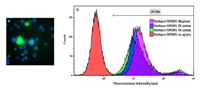
Figure 1: (a) Confocal microscopy of ALMs co-incubated
with 4% SPION solution (20×). Green denotes the presence of SPIONs and blue denotes
the nuclei of the ALMs. (b) Flow cytometry of rat ALMs co-incubated with
increasing concentrations of SPIONs. The horizontal axis displays the FL1
channel intensity.
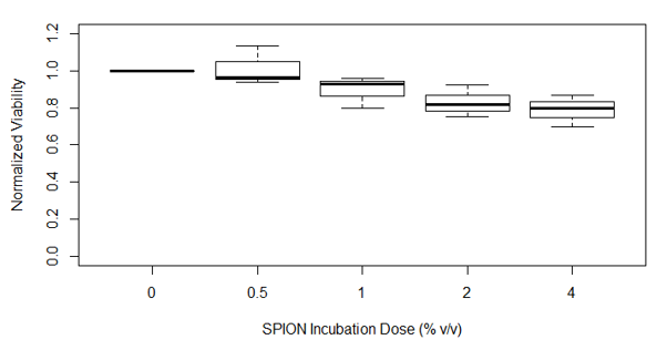
Figure 2: Viability of the labeled ALMs normalized to unlabeled
cells after four hours of incubation with increasing SPION concentrations. Cell
viability after incubation with 4% SPIONs showed a significant difference from
unlabeled cells, but still shows approximately 80% viability. Data were
analyzed using a one-way ANOVA and a Dunnet’s post-hoc test.
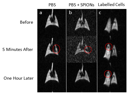
Figure 3: Images before, five minutes after and one hour
after instillation of (a) PBS, (b) PBS + SPIONs, (c) 2 million ALMs labeled
with 4% SPIONs. Areas of signal loss are circled in red. Note the persistence
of signal loss in the lungs instilled with the SPION-labeled ALMs at the
lower right.
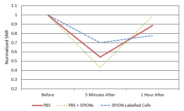
Figure 4: Normalized 129Xe SNR of regions of
interest shown by red circles in Fig. 3 before/after and one hour following
instillations. Note the persistence of the signal hypointensity for the SPION-labeled ALMs at one hour, not seen with the other installates, indicating the cells
are retained in the lungs. The amount of total SPIONs delivered with PBS and
the amount within ALMs differed, resulting in a difference of signal drop
magnitude. It is also possible that gas could not reach certain regions because
they were blocked by the instillates.