0062
Multiband zoom TSE imaging: increasing efficiency with multiband tip-back preparation pulses1School of Biomedical Engineering and Imaging Sciences, King's College London, London, United Kingdom
Synopsis
Inner volume imaging can be appealing as it negates the need to encode a large field of view when the region of interest resides within a larger tissue structure. However, conventional zoom approaches with precisely limited field of view produce strong saturation throughout the image volume, placing a restrictive lower limit on the minimum TR, in order to avoid reduced signal and contrast. Here we present a new multiband tip-back preparation pulse in combination with a zoom multiband TSE sequence, which realises the benefits of reduced field of view encoding without the penalty of saturation in the intermediate slice areas.
Introduction
Inner volume imaging typically applied alongside methods such as turbo spin-echo (TSE) or echo-planar imaging (EPI) have numerous applications where the region of interest resides within a larger tissue structure. This approach can be appealing as it negates the need to encode a large field of view which may result in long echo trains and limited resolution, particularly in the case where multi-shot acquisition degrade image quality due to motion.
A highly effective method of achieving a reduced in plane field of view uses an orthogonal excitation slab1. However, each new excited slice produces strong saturation throughout the image volume, placing a restrictive lower limit on the minimum TR, in order to avoid reduced signal and contrast of long T1 tissues.
Here we present a new multiband tip-back preparation (TBP) pulse in combination with a zoom multiband (MB) TSE sequence, which realises the benefits of reduced field of view encoding without the penalty of saturation in the intermediate slice areas.
Methods
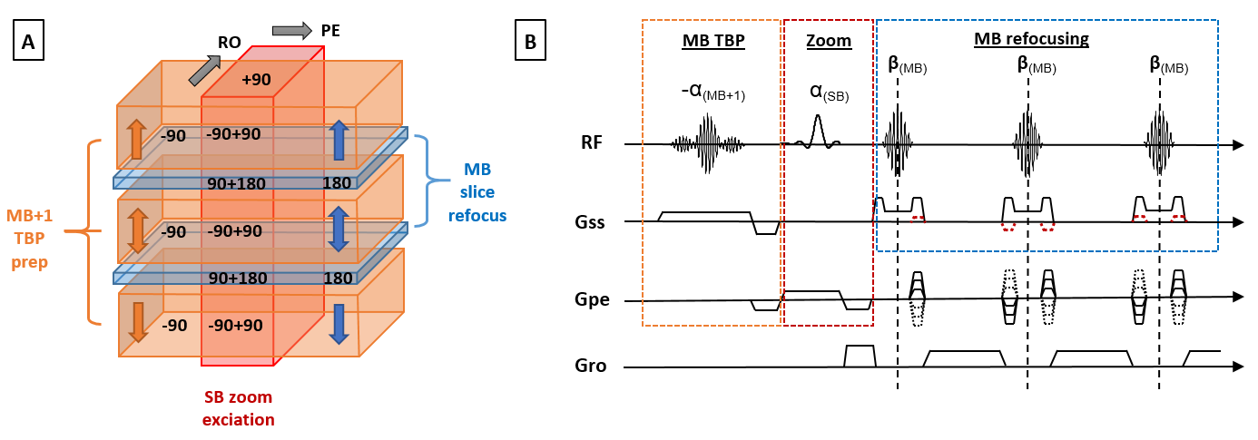
Zoom MB-TSE is implemented using a single-band slab excitation that is orthogonal to the imaging plane(s) which are imaged with multiband refocusing pulses2.The excitation pulse leads to unwanted saturation of magnetization between the imaged slices. For a sequence with MB simultaneously excited slices, the proposed multiband preparation (shown in figure 1) excites MB+1 wide slices with flip angle -90, effectively exciting the opposed pattern to the later refocusing pulses in the areas between MB slices. The zoom excitation pulse rotates the magnetization that was not affected by the prep pulse into the transverse plane, while returning the rest of the magnetization to the z-axis. Hence the magnetization between multiband excited slices remains unsaturated, meaning that a much shorter TR can be utilised. Subsequent multiband refocusing pulses are used as normal.
The method was implemented on a 3T Philips Achieva scanner using a standard MB CAIPI framework. The MB refocusing pulses operated on 2.5mm slices and the zoom excitation pulse was a wide-band, high time-bandwidth product pulse (BW: 2.3 kHz, TBP: 8.016). The tip-back preparation pulse designed to achieve a 12 mm gap around the imaging 'slices' was found effective for the desired slice thickness.
The method was tested using a 32 channel head coil when imaging an adult brain (in-plane resolution 1.5x1.5 mm, TE=120ms, refocusing pulse angle of 130) and a 32 channel cardiac coil when imaging the brain of a 27 gestational week fetus (resolution: 1.5x1.5 mm, TE=250ms, max b1 adjusted to balance SAR vs minimum echo spacing, partial Fourier factor ~0.65%). All fetal protocols operated in low SAR mode (local torso limiting whole body to ~0.8W/Kg), low PNS mode, and reduced gradient slew to limit acoustic noise).
For prostate a standard clinical multi-shot TSE sequence (multi-slice, TE=110ms, 1x1x2 mm resolution) was used alongside a single-shot zoom-TSE (single-band, TE=110ms, 1x1x2 mm resolution, Sense=2, partial Fourier=0.625) with and without MB tip-back preparation.
Written informed consent was obtained prior to scanning. Data was processed and reconstructed offline using custom unfold scripts and utilising ReconFrame (GyroTools, Zurich, CH).
Results and Discussion
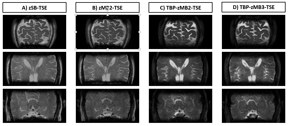
Figure 2 shows data from an adult brain using zoom TSE with various MB slice accelerations, with and without the MB tip-back preparation. The images using the preparation pulse demonstrate that much of the magnetisation has now been restored leading to enhanced signal and contrast, particularly in CSF.
Figure 3 demonstrates the improvement of the tip-back pulse for zoom TSE in the fetal brain, here it has been applied in front of MB2-TSE.
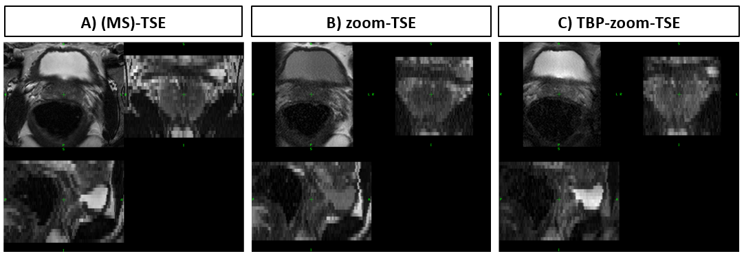
In figure 4 the method has been applied to the prostate, with a comparison to a standard clinical protocol employing multi-shot multi-slice TSE alongside zoom single-shot TSE (single band) which suffers from signal saturation at minimum TR; applying the MB TBP recovers the signal from long T1 tissue with contrast similar to the conventional multi-slice TSE image.
Conclusion
There are a number of alternative methods in use that seek to address the issue of saturation in multi-slice stack imaging whilst achieving inner volume excitation. These include 2DRF composite pulses3 and oblique 90-180 intersection to limit the extent of the saturation4. Both have been successfully applied to diffusion-EPI but may be difficult to apply alongside MB-TSE without using a long excitation pulse or limited by the jump required between consecutive imaged slices.
The proposed MB+1 tip-back pulse allows multiband zoomed TSE imaging using orthogonal slab excitation to overcome the serious limitation of between-slice saturation, allowing for much shorter TR while retaining the benefits of reduced field of view encoding. The method is also useful for single-band zoomed imaging (using a 2-band prep pulse).
Acknowledgements
This work received funding from the European Research Council under the European Union’s Seventh Framework Programme (FP7/20072013)/ERC grant agreement no. 319456 (dHCP project), and was supported by the Wellcome EPSRC Centre for Medical Engineering at Kings College London (WT 203148/Z/16/Z), MRC strategic grant MR/K006355/1 and by the National Institute for Health Research (NIHR) Biomedical Research Centre based at Guy’s and St Thomas’ NHS Foundation Trust and King’s College London. The views expressed are those of the authors and not necessarily those of the NHS, the NIHR or the Department of Health.References
[1]: Feinberg, D. Inner Volume MR Imaging: Technical Concepts and Their Applications. Radiology 1985; 156:743-747
[2]: Price, A. Multiband TSE Imaging of the Fetal Brain at 3T. Proc. Intl. Soc. Mag. Reson. Med. 24 (2016) 3249.
[3]: Finsterbusch, J. High-Resolution Diffusion Tensor Imaging with Inner Field-of-View EPI. JMRI 29:987–993 (2009).
[4]: Wheeler-Kingshott, C. ADC Mapping of the Human Optic Nerve: Increased Resolution, Coverage, and Reliability with CSF-Suppressed ZOOM-EPI. Magnetic Resonance in Medicine 47:24–31 (2002).
Figures