0022
Volumetric resonators based on novel materials for 3 T MRIAnna Mikhailovskaya1, Alena Shchelokova1, Dmitry Dobrykh1, Ivan Sushkov2, Alexey Slobozhanyuk1,3, and Andrew Webb4
1Department of Nanophotonics and Metamaterials, ITMO University, Saint Petersburg, Russian Federation, 2Department of Radiology, Vreden Russian Institute of Traumatology and Orthopedics, Saint Petersburg, Russian Federation, 3Nonlinear Physics Center, Research School of Physics and Engineering, Australian National University, Canberra, Australia, 4C.J. Gorter Center for High Field MRI, Department of Radiology, Leiden University Medical Center, Leiden, Netherlands
Synopsis
We propose and characterise a novel metamaterial-inspired approach which reduces substantially the required outer diameter of a dielectric resonator thus making possible to design compact structures for 3 T. When used in an inductively-coupled wireless mode, the sensitivity of the “meta-resonator” was measured to be slightly higher than that of a standard dielectric resonator operating in its degenerate circularly-polarized HEM11 modes. This study demonstrates the first application of a metamaterial-based approach to MR volume coil design.
Introduction
Recently, it has been shown that resonators based on high permittivity dielectric materials can be used for local and global RF shimming and local SNR increase for ultra-high frequency magnetic resonance imaging and microscopy.1-3 However, it is very difficult to design such resonators for clinical field strengths because of the relatively large geometric dimensions of structures realised with moderate permittivity. An alternative approach is to use novel structures, such as metamaterials and metasurfaces, for local sensitivity enhancement.4,5 In this study we propose a new design of a compact annular dielectric resonator based on the combination of wire metamaterials structure and high permittivity dielectric material.Methods
Figure 1 shows a schematic view of the geometries of a conventional annular dielectric resonator used for comparative experiments (a) and the new meta-resonator (b). The inner diameter d=100 mm and length h=232 mm were equal for both structures, while the outer diameters were different: dielectric resonator DDR=352 mm and meta-resonator DMR=220 mm. The permittivity of distilled water used in both resonators is 78 with a conductivity of 0.006 S/m. The wire length lw=182 mm was chosen to be approximately half of the wavelength at the operational frequency of 3 T MRI. The thickness of Plexiglas, which was used as a container for structures is 5 mm, with a permittivity ɛ=3.5. The wires were placed in such way that the spacing between them was 4 mm and the distance from the air annulus was 10 mm. The dimensions of the structures were optimized numerically using the frequency domain solver in commercially available software (CST Microwave Studio 2017). Electromagnetic simulations were performed with the annular dielectric resonator and meta-resonator where the phantom and a transmit body (birdcage geometry) coil (inner diameter 60 cm, length 213 cm) were included. We used a homogeneous cylindrical phantom with relative permittivity 81 and electrical conductivity of 0.6 S/m, a radius of 33 mm and length of 395 mm. All B1+ field and specific absorption rate (SAR) distributions were normalized to an accepted power of 1 Watt. The resonant frequency of each structure (together with the phantom placed inside the annulus) was measured using a pick-up loop (diameter 80 mm) placed above the resonator and portable vector network analyzer both outside and inside the scanner: minimal detuning was observed inside the scanner, indicating a weak coupling to the body coil. All MRI experiments were performed on a 3 Т Siemens Magnetom Verio whole-body clinical system at the Russian Scientific Research Institute of Traumatology and Orthopedics named after R.R. Vreden. Phantom images were acquired using a gradient echo sequence. Scan parameters were: field of view (FOV) 300x300 mm2, acquisition matrix 512x512, TR/TE 573/12 ms.Results
Figure 1(c) demonstrates the numerically calculated dependence of the resonant frequency of the HEM11 mode on the outer diameter of the resonators: the outer diameter of a cylindrical dielectric resonator at 123.3 MHz is 322 mm. The corresponding value for the meta-resonator is much smaller, 214 mm. Figure 2 demonstrates the results of electromagnetic simulations with the body coil transmit and cylindrical phantom placed inside the annular dielectric resonator (Figure 2(a)) and meta-resonator (Figure 2(b)). For both cases, the magnetic field localized in the central part of the resonators and highly homogeneous, while the electric field is localized outside the region of interest. Figure 3 shows gradient echo images of a courgette which was used as a test object. The body coil was used for transmission, while for receive two different configurations were used: body coil inductively coupled to the annular dielectric resonator (Figure 3(a)) and body coil inductively coupled to the meta-resonator (Figure 3(b)).Discussion
A new meta-resonator design supports the eigenmode profile which is similar to the HEM11 mode of the dielectric resonator at the desired frequency while having dimensions much smaller than the conventional annular dielectric resonator. Both the resonators significantly increase the SNR.Conclusions
We have proposed and tested a new design of annular resonator based on the combination of a wire metamaterial-inspired structure and high permittivity dielectric material. The meta-resonator acts as a passive structure which electromagnetically couples with the transmit body coil to produce a very homogeneous B1+ field distribution in the region-of-interest. While the performance of the annular dielectric and meta-resonators are almost identical the geometrical dimensions differ significantly which is crucially important for practical applications.Acknowledgements
This research was supported by Ministry of Education and Science of the Russian Federation (Zadanie No. 3.2465.2017/4.6). The authors are grateful to Dr. M. Zubkov and to Mr. E. Kretov for assistance with measurements and to Dr. C. A. T. van den Berg, Dr. A. J. Raaijmakers, Dr. I. V. Melchakova, Prof. Y. S. Kivshar and Prof. P. A. Belov for useful discussions at the preliminary stage of the project.References
1. Aussenhofer S A and Webb A G. Design and Evaluation of a Detunable Water-Based Quadrature HEM11 Mode Dielectric Resonator as a New Type of Volume Coil for High field MRI, Magn Reson Med. 2012; 68:1325-1331. 2. Aussenhofer S A and Webb A G. High-permittivity solid ceramic resonators for high-field human MRI. NMR Biomed. 2013; 26:1555. 3. Bluemink J J, et al. Dielectric wave-guides for ultrahigh field magnetic resonance imaging. Magn. Reson. Med. 2016; 76: 1314–1324. 4. Sobozhanyuk A P, et al. Enhancement of Magnetic Resonance Imaging with Metasurfaces. Adv. Mater. 2016; 28:1832–1838. 5. Schmidt R, et al. Flexible and compact hybrid metasurfaces for enhanced ultra high field in vivo magnetic resonance imaging. Sci. Rep. 2017; 7: 1678.Figures
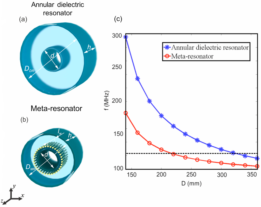
Figure 1. Schematic view of the
geometries of the annular dielectric resonator (a) and meta-resonator (b). The inner diameter d=100 mm and the height h=232
mm are equal for both structures, while the outer diameters are different:
dielectric resonator DDR=352
mm and meta-resonator DMR=220
mm. The length of wires lw=182
mm and the thickness of plexiglass is 5
mm. (с) Numerically calculated resonance frequency of
the HEM11 eigenmode as a function of the outer diameter (D) for an
annular dielectric resonator (blue curve) and meta-resonator (red curve),
dashed black curve indicates the Larmor frequency at 3 T.
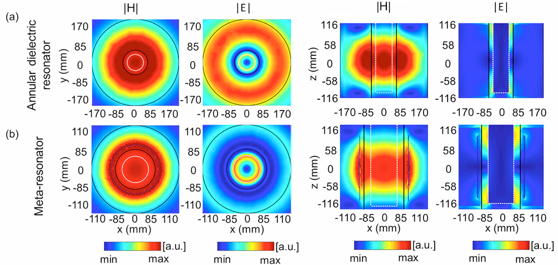
Figure 2. Numerically
calculated magnetic (|H|) and electric
(|E|) fields amplitudes in
yx- and zx-planes for an annular dielectric resonator (a) and
meta-resonator (b) with a homogeneous cylindrical phantom imitating human hand placed
inside. White dashed lines indicate the boundaries of the phantom, black solid lines
show the resonators boundaries.
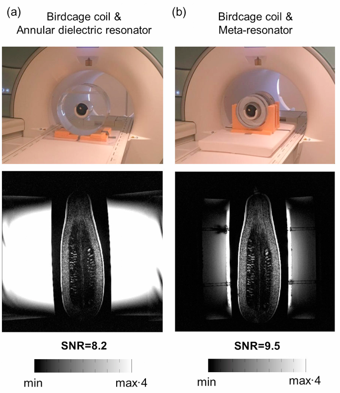
Figure 3. Photographs
of the experimental setups and MR images of a courgette obtained (a) with the birdcage coil and annular
dielectric resonator, (b) with the birdcage coil and meta-resonator.