0016
The iPRES-W AIR Coil: A Flexible RF Coil for Simultaneous MR Image Acquisition, Wireless Communication, and Localized B0 Shimming1Brain Imaging Analysis Center, Duke University, Durham, NC, United States, 2Medical Physics Graduate Program, Duke University, Durham, NC, United States, 3GE Healthcare, Aurora, OH, United States
Synopsis
The iPRES-W AIR coil is a highly novel RF coil design that can simultaneously perform image acquisition, wireless communication, and wirelessly controlled localized B0 shimming. In addition, the iPRES-W AIR coil benefits from all advantages of the recently unveiled AIR coil technology, which offers a flexible and ultra-lightweight coil for increased patient comfort and freedom in overlap positioning between coil elements without degrading the performance. This technology has enormous potential to improve image quality, spatial fidelity, diagnostic accuracy, and patient comfort in a wide range of MRI applications.
Introduction
RF coil design has become increasingly complex from an MR physics and electronics perspective. This complexity manifests itself in a clinical environment by the large number of coils required for different applications. These coils are typically bulky, heavy, and rigid, have many wired connections, and include specialized coils such as localized shim coil arrays1. Here, we propose a highly innovative coil design that can simultaneously perform MR image acquisition, wireless communication, and localized B0 shimming, while also being flexible and ultra-lightweight.
First, our coil design implements the concept of integrated parallel reception, excitation, and shimming (iPRES)2, which enables simultaneous imaging and localized B0 shimming with a single coil array without compromising the signal-to-noise ratio (SNR). Second, it integrates a novel technology that further enables wireless communication with the same coil array to minimize the number of wired connections for data transfer between the coil array and the scanner3. Finally, it benefits from all advantages of the recently unveiled AIR coil technology, which provides not only flexible and ultra-lightweight coils for increased patient comfort, but also more flexible coil design opportunities resulting from the ability to optimize the overlap between coil elements without degrading the performance4,5.
Methods
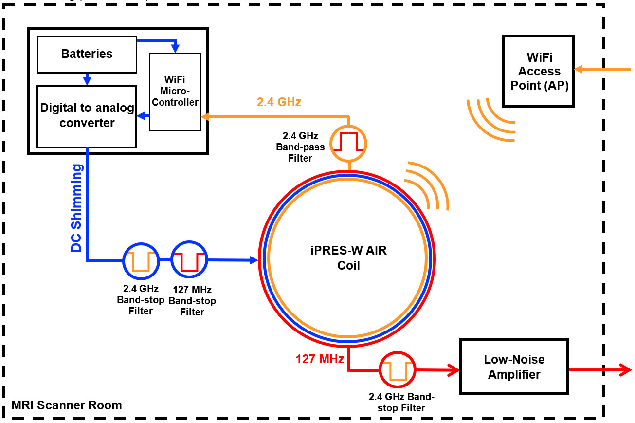
As a proof-of-concept, a single 11-cm AIR coil was modified to enable three independent currents to flow in the loop: an RF current at the Larmor frequency (127 MHz for a 3T scanner) for MR imaging, an RF current at 2.4 GHz for WiFi communication, and an adjustable DC current for localized B0 shimming. To ensure that the two RF transmission lines were isolated from one another, a band-pass filter and an LC band-stop filter were integrated in series with the WiFi and MRI feed lines, respectively (Fig. 1). Specifically, the band-pass filter between the onboard WiFi microcontroller and the RF coil provided -30 dB of isolation at 127 MHz, while the band-stop filter between the MRI port and the RF coil provided -20 dB of isolation at 2.4 GHz. Proper isolation ensured no deterioration of image SNR or WiFi signal strength and protection from unwanted currents damaging onboard circuit components.
Implementing iPRES to add the DC current required the same RF protection on the twisted-pair DC power cable, using both 127 MHz and 2.4 GHz band-stop filters for isolation. The internal layout of the AIR coil provided protection from the DC current reaching the two RF transmission lines. To provide the adjustable DC currents without using additional wired connections, a 4-channel circuit powered by MR-compatible batteries converted the digital pulse-width-modulated output of the WiFi microcontroller to an analog output. Each channel supplied an output current with a range of ±1 A and a resolution of 2 mA. The current values were updated from outside the scanner room using the onboard wireless communication, allowing for remotely controlled shimming.
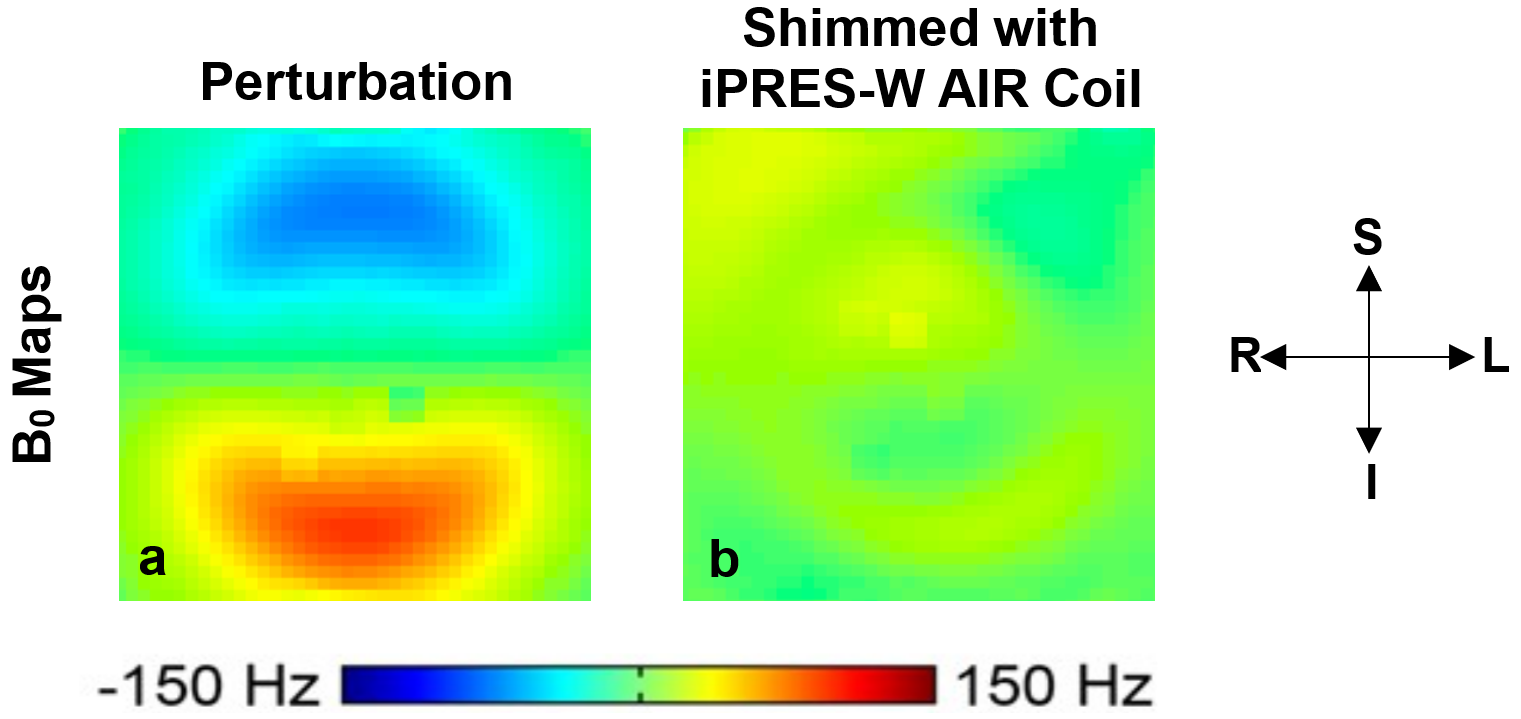
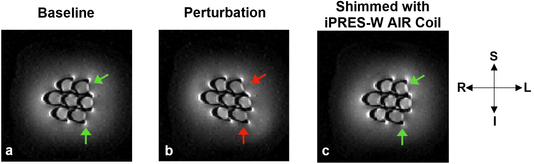
Proof-of-concept experiments were performed on a 3T scanner to show the ability of this new iPRES-W AIR coil to shim using wireless communication. A DC current was applied in a perturbation loop placed on a water phantom to create a localized B0 inhomogeneity. The optimal DC current to shim each slice was calculated as described previously2 and wirelessly sent to the coil. Coronal B0 maps and EPI images were acquired before and after shimming. In addition, the WiFi performance of the iPRES-W AIR coil was monitored by measuring and comparing its received signal strength indicator (RSSI), at incremental distances from an access point (AP), with that of the unmodified onboard antenna of the WiFi microcontroller.
Results
Wirelessly shimming with the iPRES-W AIR coil drastically reduced the perturbation-induced B0 inhomogeneities (Fig. 2) and geometric distortions in the EPI images (Fig. 3). Its WiFi Performance showed an average RSSI improvement of 3 dB in comparison to the onboard antenna of the microcontroller.Discussion and Conclusion
These proof-of-concept experiments demonstrate the effectiveness of our new highly flexible and ultra-lightweight iPRES-W AIR coil at performing simultaneous image acquisition, WiFi communication, and localized B0 shimming. By using battery-supplied DC shim currents, we further progress towards the ability to remove all wired connections between the scanner and the coil. Further applications can use wireless communication not only for remote-controlled shimming, but also for other types of data transfer between the coil and the scanner. This highly innovative technology, which integrates three different functions into a single flexible coil, represents a paradigm shift from the traditionally large and bulky coils used in a clinical environment and has enormous potential for improving image quality, spatial fidelity, diagnostic accuracy, and patient comfort in a wide range of MRI applications.Acknowledgements
This work was in part supported by grants R21 EB018951, R24 MH106048, R21 EB024121 from the National Institutes of Health, by GE Healthcare, and by the Duke-Coulter Translational Partnership.References
1. Juchem C et al. J Magn Reson 2011;212:280-8.
2. Truong TK et al. NeuroImage 2014;103;235-40
3. Darnell D et al. WiFi-enabled RF Coil for Simultaneous MR Image Acquisition and Wireless Communication. Proceedings of the ISMRM, April 2017, Honolulu: pg. no. 4430.
4. Stormont R et al. Reimagining Flexible Coil Technology. GE SIGNA Pulse of MR, V. 21, Spring 2017
5. Vasanawala S et al. Development and Clinical Implementation of Very Light Weight and Highly Flexible AIR Technology Arrays. Proceedings of the ISMRM, April 2017, Honolulu: pg. no. 0755.
Figures