0014
Dynamic flow imaging and quantification using cine FISS arterial spin labelingRobert R Edelman1,2, Ali Serhal2, Amit Pursnani3, Jianing Pang4, and Ioannis Koktzoglou1,5
1Radiology, NorthShore University HealthSystem, Evanston, IL, United States, 2Radiology, Feinberg School of Medicine, Northwestern University, Chicago, IL, United States, 3Medicine, NorthShore University HealthSystem, Evanston, IL, United States, 4Siemens Medical Systems, Chicago, IL, United States, 5Radiology, Prtizker School of Medicine, University of Chicago, Chicago, IL, United States
Synopsis
We describe a new approach for flow imaging and quantification consisting of a prototype cine arterial spin labeling (ASL) pulse sequence using a highly-accelerated radial fast interrupted steady-state (FISS) readout. The technique was successfully applied in several vascular regions (coronary arteries, pulmonary arteries, renal arteries, circle of Willis). These preliminary results suggest that cine FISS ASL has the potential to provide an efficient and visually-appealing alternative to phase contrast for the depiction and quantification of blood flow.
Introduction
We describe a new approach for flow imaging consisting of a prototype cine arterial spin labeling (ASL) pulse sequence using a highly-accelerated radial fast interrupted steady-state (FISS) readout. Unlike previously described ASL techniques, the new approach is both efficient and quantitative, enabling the straightforward measurement of flow velocity.Methods
Validation of the cine FISS ASL technique was initially performed in a pulsatile flow phantom. An IRB-approved study was then conducted in healthy volunteers on a 1.5 Tesla scanner (MAGNETOM Avanto, Siemens Healthcare, Erlangen, Germany). The technique was evaluated in several vascular territories (coronary, pulmonary and renal arteries, and the circle of Willis). The FISS readout [1] differs significantly from a conventional bSSFP readout in that the steady-state magnetization undergoes gradient and RF spoiling after each block of 6-8 bSSFP modules. A radial k-space trajectory with equidistant azimuthal view angles suppresses image artifacts that would otherwise occur due to the periodic disruption of the steady-state signal. Scan parameters included: slice thickness = 2 to 12-mm, radial views = 96 to 192, acquisition matrix = 144, 8 shots, sampling bandwidth ~ 890 Hz/pixel; retrospective electrocardiographic and/or pulse gating. Inflowing spins were labeled using a 25 to 50-mm thick adiabatic inversion radiofrequency (RF) pulse. Two cine FISS ASL approaches were tested: (1) a “standard” approach, where background suppression was obtained by complex subtraction of two interleaved cine image series, one with RF labeling of inflowing spins and one without; (2) a “self-subtractive” approach, where only one set of images was acquired, and background suppression was obtained by complex subtraction of a late cine frame from all cine frames. Except for the circle of Willis, cine FISS ASL scans were acquired in a single breath-hold with scan time of 16 heartbeats (standard approach) or 8 heartbeats (self-subtractive approach). The instantaneous flow velocity was quantified as the ratio of: (distance traveled by the leading edge of the labeled blood) / (frame duration).Results
The flow phantom study showed excellent correlation (r2 = 0.9974, p = 0.001) between cine FISS ASL and phase contrast measurements of flow velocity. In seven volunteers in whom the standard technique was assessed in the coronary arteries, cine FISS ASL showed smooth progression of the tagged spins through the left main and left anterior descending coronary arteries (Figure 1A). Mean coronary flow velocity, measured over an ≈209 ± 97 msec (mean ± sd) span of diastole was 11.7 ± 3.0 cm/sec. In addition, the technique proved reliable for visualizing and quantifying flow outside of the heart (Figures 1B and 1C). For extra-cardiac regions, image quality was similar for both cine FISS ASL techniques despite the two-fold reduction in scan time using the self-subtractive approach.Discussion and Conclusion
These preliminary results suggest that cine FISS ASL has the potential to provide an efficient and visually-appealing alternative to phase contrast for the depiction and quantification of blood flow. The use of a FISS readout provides several benefits, including: (1) intrinsic fat suppression which minimizes streak artifacts from the use of a highly accelerated radial acquisition; (2) suppression of image artifacts that would otherwise result from disruption of the steady-state magnetization each time the RF labeling pulse is applied; and (3) reduction of flow artifacts. Compared with 2D phase contrast, the method offers several potential benefits: (1) it is unaffected by background phase shifts; (2) the signal-to-noise ratio is greatly enhanced using a steady-state rather than spoiled gradient-echo readout; and (3) for imaging of in-plane flow, there is negligible flow-related saturation. Moreover, the near-perfect degree of background suppression facilitates the use of thick slices for semi-projective imaging of in-plane flow, without the partial volume averaging encountered with 2D phase contrast.Acknowledgements
NIH grants R01 HL130093 and R21 HL126015.References
1. Koktzoglou I, Edelman RR. Radial fast interrupted steady-state (FISS) magnetic resonance imaging. Magn Reson Med. 2017 Aug 30. doi: 10.1002/mrm.26881. [Epub ahead of print]Figures
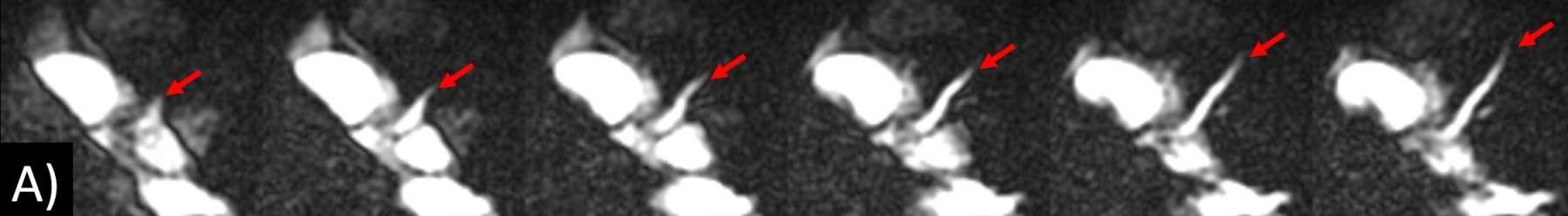
1. Cine FISS ASL applied
to different vascular territories (six frames shown out of 32 acquired). (A) Imaging of the left main and left
anterior descending coronary artery (arrows) in an oblique axial plane.
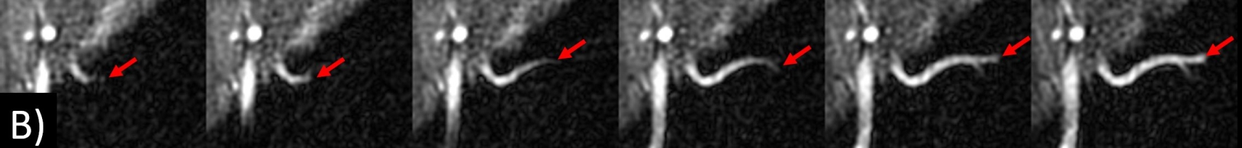
1B. Cine FISS ASL of the left renal artery (arrows) in an oblique coronal plane.
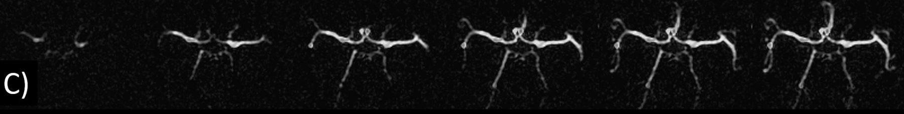
1C. Cine FISS ASL
of the circle of Willis.