MR-Guided Radiotherapy
1UMC Utrecht, Netherlands
Synopsis
Online MRI guidance is the new disruptive technology for radiotherapy that will facilitate online and real-time adaptive treatments. An overview of the current hybrid MRI-guided treatment machines will be given. The MRI-Linac, which combines a 1.5T closed bore system with a modern 7MV linear accelerator will be described. Its clinical introduction is highlighted and the potential for future treatments and research is outlined.
Target Audience
Clinicians interested in learning about the clinical potential of the MRI-Linac. Scientists who want to know more about the new research field that is being created by the introduction of the MR-Linac.Highlights
· Online MRI-guidance is a disruptive new technology that will completely overhaul the radiotherapy process.
· Several hybrid MRI-guided radiotherapy systems are currently being developed. Each with its unique choices in design.
· Radiotherapy imaging requirements are different compared to radiological applications as well as the imaging hardware. For this purpose novel MR methods for real-time guidance are being developed.
· MRI-guided radiotherapy will open up a whole new field of research in the coming years ranging from methods development (e.g., real-time imaging) to clinical research (e.g., tumor response modelling) and data science.
Introduction
The use of MRI in radiotherapy is growing rapidly. MRI for radiotherapy treatment planning, where MRI data is used to aid the delineation of tumor and organs at risk, is becoming the standard for many tumor sites. The integration of MRI in the radiotherapy workflow is motivated by the superior soft tissue contrast as compared to CT. Radiotherapy has the unique ability to deliver a differential treatment. Modern day treatment machines (i.e., linear accelerators, LINACs) are able to deliver complex dose distributions to the target volume. For this reason, there has been considerable research effort to combine PET imaging with multi-parametric MRI (e.g., T1, T2, DWI and DCE information) to characterize the biological properties within the tumor and use this information to escalate the dose to parts of the tumor that are aggressive or radioresistent (van der Heide et al. 2012, Lagendijk et al. 2014).Imaging Requirements and Challenges
Implementing MRI in a
radiotherapy workflow, however, brings new challenges. Current Radiotherapy
treatments are given in multiple fractions that are based on the same reference
image. In current day radiotherapy this is a CT image that provides an attenuation
map needed to optimize the radiation delivery (i.e., treatment planning). For
this reason all preparatory imaging, including MRI, is performed in radiotherapy
treatment position. The requirement for a flat table top and positioning accessories,
such as arm supports and thermoplastic masks, often limits optimal coil placement,
which has a direct effect on SNR. Moreover, the geometric accuracy requirements
are much higher for radiotherapy compared to radiological applications.The MRI Linac
The holy grail of image-guided
radiotherapy is to see the tumor while
the patient is being irradiated. With current CT based onboard imaging this is
not possible. In 2000 Lagendijk and Bakker proposed the use of a hybrid MRI-Linac
system for online and real-time guidance of the treatment. Several integrated
systems with varying field-strengths and magnet designs are now under
development (Lagendijk et al. 2008,ViewRay®, Fallone et al. 2009, Constantin et
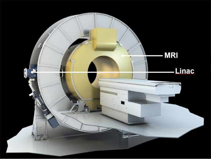
al. 2011). At the UCMU, a hybrid 1.5T closed-bore MRI with 7 MV Linac is being
developed in collaboration with Elekta and Philips (Fig. 1). To avoid magnetic coupling
between the accelerator and the MRI system, the active shielding was modified
to create a zero magnetic field in a toroid closely around the magnet (Overweg
et al. 2009). The Faraday cage has been redesigning to prevent RF interference
from the Linac’s microwave, and special radiolucent RF receiver coils were built
to minimize attenuation of the treatment beam. It was shown with an
experimental prototype (Raaymakers et al. 2008) that both systems fully
function and work completely independent.Clinical Introduction of MR-Guided Systems
In 2014 the first patient has
been treated on a Viewray MRIdian 0.35T split-magnet with Cobalt radiation
sources. Currently patients are being treated on this machine in six treatment
centers world wide. Elekta is currently installing pre-clinical prototypes in
seven centers, which form an international consortium that collaborates on the clinical
introduction of the MRI Linac. Within this consortium predicate studies have been
conducted and clinical trials are being designed to safely introduce this new
technology into the clinic and assess its effectiveness following the IDEAL recommendations
(McCulloch et al. 2009). Within the UMC Utrecht, we are currently preparing for
the first in man experiment.New (MRI) Technology needed
MRI-guided radiotherapy will open up a whole new field of research in the coming years: having patients treated multiple times in the MRI over the course of several days or weeks will provide a unique opportunity to develop accurate models for tumor response monitoring, NTCP modelling, and dose escalation studies. However, to provide these studies with the best possible imaging data novel methods are needed such as low-field functional MRI methods to characterise the tumor response, and dynamic (3D) real-time imaging to accurately track the dose deposition (Stemkens et al. 2016). All these methods will have to work within the constraints of hybrid systems and provide exquisite geometric accuracy. Finally, with this new approach of MR-guided radiotherapy enormous amounts of multi-dimensional MRI data will be generated, so data science will become increasingly important in the future.Acknowledgements
No acknowledgement found.References
1. Constantin DE, Fahrig R, Keall PJ. A study of the effect of in-line and perpendicular magnetic fields on beam characteristics of electron guns in medical linear accelerators. Med Phys. 2011; 38:4174-85
2. Fallone BG, Murray B, Rathee S, Stanescu T, Steciw S, Vidakovic S, Blosser E, Tymofichuk D. First MR images obtained during megavoltage photon irradiation from a prototype integrated linac-MR system. Med Phys. 2009; 36:2084-8
3. Van der Heide UA, Houweling AC, Groenendaal G, Beets-Tan RGH, Lambin P. Functional MRI for radiotherapy dose painting. MRI. 2012; 30(9):1216-23
4. Lagendijk JJW and Bakker CJG, MRI guided Radiotherapy, a MRI based linear accelerator. ESTRO Istanbul 2000
5. Lagendijk JJW, Raaymakers BW, Van den Berg CAT, Moerland MA, Philippens ME, van Vulpen M. MR guidance in radiotherapy. Phys Med Biol. 2014; 59(21):349-69
6. Lagendijk JJW, Raaymakers BW, Raaijmakers AJE, Overweg J, Brown KJ, Kerkhof EM, van der Put RW, Hårdemark B, van Vulpen M, van der Heide UA. MRI/linac integration. Radiother Oncol. 2008; 86:25-9
7. McCulloch P, Altman DG, Campbell BC, Flum DR, Glasziou P, Marshall JC, Nicholl J. No surgical innovation without evaluation: the IDEAL recommendations. Lancet 2009; 374:1105-12
8. Overweg , Raaymakers BW, Lagendijk JJW, Brown K. System for MRI guided Radiotherapy. Proc. Intl. Soc. Mag. Reson. Med. 2009
9. Raaymakers BW, Raaijmakers AJE, Lagendijk JJW. Feasibility of MRI guided proton therapy: magnetic field dose effects. Phys Med Biol. 2008; 53:5615-22
10. Stemkens B, Tijssen RH, de Senneville BD, Lagendijk JJW, van den Berg CAT. Image-driven, model-based 3D abdominal motion estimation for MR-guided radiotherapy. Phys Med Biol. 2016: 61(14):5335-55
Figures