Imaging Applications of Ferumoxytol for MRI: Focusing on the Vasculature & Inflammation
Edward Neuwelt1 and Gerda Toth1
1Oregon Health & Science University, Portland, OR, United States
Synopsis
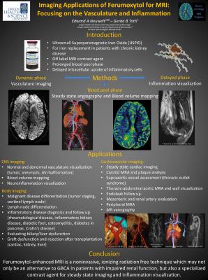
Ferumoxytol is an ultrasmall, paramagnetic iron oxide and also a novel magnetic resonance imaging (MRI) contrast agent. With its unique features (long plasma half-life and delayed intracellular uptake) ferumoxytol may pay a crucial role in the MR imaging in the future. We have completed over 700 MRI studies with ferumoxytol in our institution, primarily for CNS imaging. In this presentation we go through the general properties and the specific opportunities of ferumoxytol-enhanced MRI in and outside the brain.
Imaging Applications of Ferumoxytol for MRI: Focusing on the Vasculature and Inflammation
Ferumoxytol, a super paramagnetic iron oxide nanoparticle that is FDA approved for iron replacement therapy, is a novel magnetic resonance imaging (MRI) contrast agent that is showing great promise for MRI throughout the body. Furthermore, ferumoxytol is safe to use in patients with impaired renal function, unlike gadolinium and other iodinated contrast agents. With a prolonged blood pool phase, ferumoxytol nanoparticles are trapped in the vasculature as a blood-pool agent and allow for accurate perfusion measurements of blood volume. At later time points, ferumoxytol uptake by macrophages act as a biomarker for inflammation. Preclinical and clinical studies using ferumoxytol as a contrast agent, both in and outside of the CNS, are elucidating the effectiveness of this nanoparticle as a contrast agent throughout the body. Our focus will be on the approximately 630 infusions of ferumoxytol we have done at our institution, primarily for imaging CNS disease, and review the application of ferumoxytol in imaging non-CNS diseases.