Hyperpolarized 13C: New Kind on the Block
1Physical Therapy and Rehabilitation Science, UCSF, San Francisco, CA, United States, 2Radiology and Biomedical Imaging, UCSF, San Francisco, CA, United States
Synopsis
The goal of this presentation is to critically present the potential and limitations of hyperpolarized 13C Magnetic Resonance Spectroscopic Imaging (HP 13C MRSI) for diagnosis and monitoring of non-cancer neurological disorders. We will present in vivo studies in which hyperpolarized [1-13C] pyruvate was used to monitor neuroinflammation in preclinical models of Multiple Sclerosis and Traumatic Brain Injury, and discuss the results. We will also give an overview on how to design hyperpolarized 13C probes for metabolic imaging, and discuss the technical and biological requirements of such imaging agents with a special focus on brain.
HIGHLIGHTS
- New kind on the block: Hyperpolarized 13C MR metabolic imaging applied to the study of neurological disorders
- Example of applications to Multiple Sclerosis and Traumatic Brain Injury
- One step further: Introduction on how to design new hyperpolarized 13C probes for metabolic imaging
TARGET AUDIENCE
Researchers and clinicians interested in hyperpolarized 13C MR metabolic imaging and its potential for non-cancer neurological applications.OUTCOME/OBJECTIVES
Upon completion of this course, learners should have an understanding of the potential and limitations of using hyperpolarized 13C MR metabolic imaging for the study of neurological disorders. Furthermore, learners should understand the basic requirements of designing hyperpolarized 13C probes for metabolic imaging, especially in the context of brain applications.PURPOSE
The purpose of this talk is to address the following questions: “Can we use hyperpolarized 13C MR metabolic imaging to study neurological disorders? What is the potential of this method? What are the limitations? Which new probes do we need?”METHODS
This talk will refer to the following technical and biological methods/concepts:
- Dynamic Nuclear Polarization
- Hyperpolarized 13C MR metabolic imaging
- MR spectroscopic imaging
- Biochemical validation
- Preclinical models of neurological disorders
RESULTS
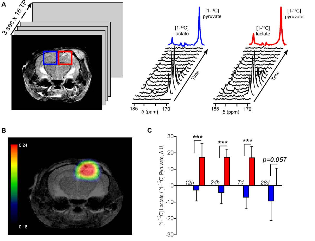
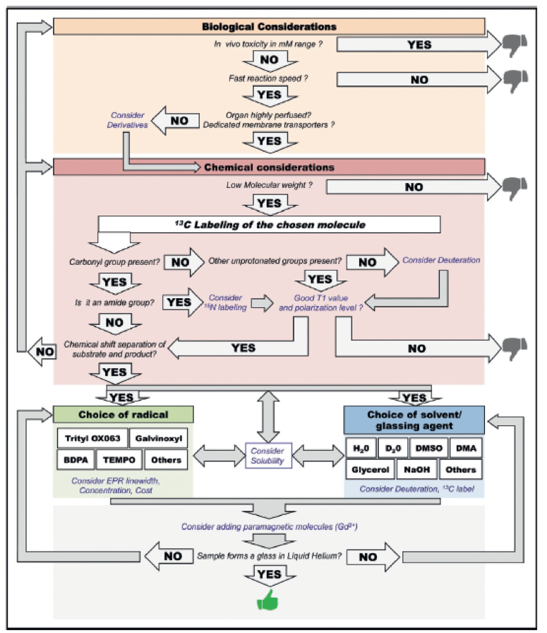
In this seminar, I will first present applications of the use of metabolic imaging of hyperpolarized [1-13C] pyruvate to the study of preclinical animal models of Multiple Sclerosis (MS) and Traumatic Brain injury (TBI) (see Abstract #4922 and #5160). Our results show that this imaging strategy detect increased hyperpolarized lactate production in vivo in highly inflammatory white matter lesions (MS model) and in impact lesions (TBI model). In MS, increased lactate production was associated with the presence of pro-inflammatory macrophages upregulating pyruvate dehydrogenase kinase-1, as well as regional inhibition of pyruvate dehydrogenase, providing a likely mechanism for a decrease subsequent flux of pyruvate towards the Krebs cycle. In TBI on the other hand, increased lactate production was associated with the presence of pro-inflammatory macrophages and PDH inhibition, but PDK-1 seemed to not be a player. Finally, in the last part on this talk, I will present the development of a new probe, namely [Guanidino-13C]-arginine, and its potential for detection of inflammatory processes. I will also give an overview on how to design a hyperpolarized probe for metabolic imaging.CONCLUSION
Because HP 13C MRSI is clinically translatable and expanding rapidly, studying the potential of this method for diagnostic and monitoring of non-cancer diseases, in particular neurological disorders, is of high significance. Preclinical validations are at this stage still scientifically needed for future clinical trials on non-cancer pathologies.Acknowledgements
This work was supported by research grants NMSS_PP3395, Cal-BRAIN349087, UCSF_RAP7500634, UCSF Department of Radiology seed grants #14-04 & #14-05, NIH R01CA172845, NIH R01NS102156, NIH Hyperpolarized MRI Technology Resource Center #P41EB013598, fellowships from the Flemish Institute for Science and Technology (IWT) and the NMSS (FG-1507-05297), and by FISM-Fondazione Italiana Sclerosi Multipla Senior Research Fellowship Cod. 2014/B/1 to AD.References
ISMRM abstracts at this conference:
Abstract #5160: Guglielmetti C, Najac C, Van der Linden A, Ronen SM, Chaumeil MM, “Metabolic imaging of neuroinflammation in the cuprizone mouse model for Multiple Sclerosis using hyperpolarized [1-13C] pyruvate”
Session: Hyperpolarized 13C Magnetic Resonance Imaging & Spectroscopy Day/Date: Monday, April 24, 2017 Session Time: 13:45
Abstract #4922: Guglielmetti C, Chou A, Krukowski K, Paladini MS, Riparip K, Rosi S, Chaumeil M "In vivo metabolic imaging of neuroinflammation following traumatic brain injury using hyperpolarized [1-13C] pyruvate"
Session: Pitch: New Molecular & Metabolic Imaging Approaches Day/Date: Tuesday, April 25, 2017 Session Time: 16:15
[1] Galvan-Pena S & O'Neill LA (2014) Metabolic reprograming in macrophage polarization. Front Immunol 5:420.
[2] Tannahill GM, Iraci N, Gaude E, Frezza C, & Pluchino S (2015) Metabolic reprograming of mononuclear phagocytes in progressive multiple sclerosis. Frontiers in immunology 6:106.
[3] Kurhanewicz J, et al. (2011) Analysis of cancer metabolism by imaging hyperpolarized nuclei: prospects for translation to clinical research. Neoplasia 13(2):81-97.
[4] Kipp, Acta Neuropathol. (2009)
[5] Chaumeil MM, Najac C, & Ronen SM (2015) Studies of Metabolism Using (13)C MRS of Hyperpolarized Probes. Methods in enzymology 561:1-71.
[6] Najac, C., Chaumeil, M.M. et al. Detection of inflammatory cell function using 13C MRS of hyperpolarized 13C-labeled arginine. Scientific Reports 6:31397 (2016).
Figures