Conventional MRI: What We are Missing
1Center for Magnetic Resonance Research, University of Illinois at Chicago, Chicago, IL, United States
Synopsis
Metabolic MR imaging at ultrahigh field using MR signals such as sodium (23Na) and oxygen (17O) advances medical imaging beyond the current paradigm of anatomical description to metabolic bioscales. Diseases with long prodromes, including neurodegenerative diseases such as Alzheimer's disease and musculoskeletal diseases such as osteoarthritis, will be reviewed for human applications. Sensitive detection of early pathology prior to anatomic changes allows these quantitative parameters to serve as objective outcome measures for evaluating clinical trials of early interventions. Eliminating the need for subjective outcome measures of clinical disease severity would revolutionize drug discovery for early intervention.
TITLE
Conventional MRI: What We are MissingTarget Audience
Radiologists and MR imaging scientists wishing to advance MR imaging beyond the current paradigm of descriptive anatomic images of late disease to quantitative metabolic imaging that can detect early disease and monitor its progression objectively.Outcome/Objectives
The increased sensitivity of ultrahigh field MR imaging can change the medical imaging paradigm from descriptive anatomic MR imaging using proton MR signals from water and fat to quantitative metabolic bioscales based on non-proton MR signals such as sodium and oxygen. These newly accessible signals, if available clinically, potentially address unmet needs in medicine in which anatomic correlates either do not exist or are late findings. By addressing early diagnosis and monitoring disease progression, such new imaging technology offers the potential to improve outcome by promoting the discovery of earlier lower cost interventions.PURPOSE
Metabolic imaging has been proposed for as long as proton MR imaging but has been handicapped by the lack of intrinsic MR sensitivity and low metabolic concentrations of the non-proton nuclei. The quadrupolar behavior of many of these nuclei with rapid biexponential relaxation behavior also presented challenges for quantitative imaging. Ultrahigh field scanners for humans at 7T, 9.4T and , more recently 10.5T, now address these challenges making timely a review of how these imaging procedures can address the current unmet needs in medicine.METHODS
Review of the ultrahigh field (>3T) literature on human imaging has indicated a number of non-proton MR signals that can be imaged quantitatively and can be essential tools for the discovery of interventions in the early stages of economically important diseases within and outside the brain. The biologically endogenous nuclei that have exploited the increased sensitivity of ultrahigh field for human imaging include sodium, oxygen, phosphorus, chloride and potassium. Quantification of these MR signals are limited to sodium and oxygen. This review will use sodium imaging as the prototype for biological measurements.RESULTS
The paradigm shift from proton anatomical MR imaging to non-proton quantitative metabolic MR imaging will be discussed using sodium MR imaging as the prototype nucleus for diseases of the brain and cartilage.DISCUSSION
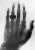
Since the first medical radiograph (Figure 1) of his spouse's hand performed by Wilhelm Conrad Roentgen in 1985, medicine has sought to investigate the human body and its anatomical changes caused by disease. This anatomical approach has brought many significant advances in treatment of disease. Yet, there are many important diseases without anatomical correlates or for which the anatomical findings appear very late in the disease thereby precluding timely diagnosis and early intervention. The neurodegenerative diseases represent an increasingly important group of diseases with long prodromal periods of decades in which a progressive pathology remains disguised by the adaptive neuroplasticity of the brain until its ultimate clinical presentation as a devastating disease with no successful treatment. Alzheimer's disease (AD) is the major example that in our aging population imposes an enormous cost in terms of quality of life for patients and their caretakers as well as an economic burden on the healthcare system (US$226 Billion in 2015). Pharmaceutical companies have focused on developing drugs attempting to halt disease progression using cognitive compromise as outcome measures in clinical trials. This has been a futile effort with no significant effective treatments. Slowly progressive diseases with long prodromes are alos common outside the brain, of which osteoarthritis is an important example in the musculoskeletal system compromising quality of life of patients and burdening the healthcare system (US$185 Billion in 2007). The prevention of early cartilage degradation has the potential to halt progression to long-term joint destruction that requires expensive joint replacement surgery to maintain mobility and quality of life. Such experiences indicate that there is an unmet need in medicine to detect and monitor diseases at its earliest stages before clinical manifestation
CONCLUSION
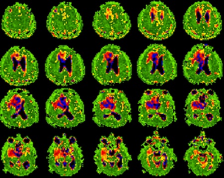
Certain capabilities are required for imaging to be useful in the discovery process of such early interventions. The first is sensitivity to the earliest stages of the disease that cannot be based on anatomy but rather on metabolic disturbances. The second is that the imaging parameters must quantitatively measure the progression of the disease. A third practical feature would be that the imaging approach can generalize across many diseases in the brain and body. Ultrahigh field MR imaging of sodium and oxygen (as well as other nuclei) can quantitatively determine cell volume fraction (CVF) (Figure 2) and metabolic rate of oxygen consumption (MRO2) and will be discussed in terms of early pathology detection and intervention discovery.Acknowledgements
The author acknowledges the support of NIH grant RO1 NS0959745-01A1.References
1. Thulborn KR. Quantitative sodium MR imaging: A review of its evolving contribution to medicine. Neuroimage 2016; 29(2):137-43.
2. Thulborn et al., Quantitative sodium MRI in the human brain at 9.4T provides assessment of tissue sodium concentration and cell volume fraction during normal aging, NMR Biomed 2016;29(2):137-43.
Figures