Renal
Synopsis
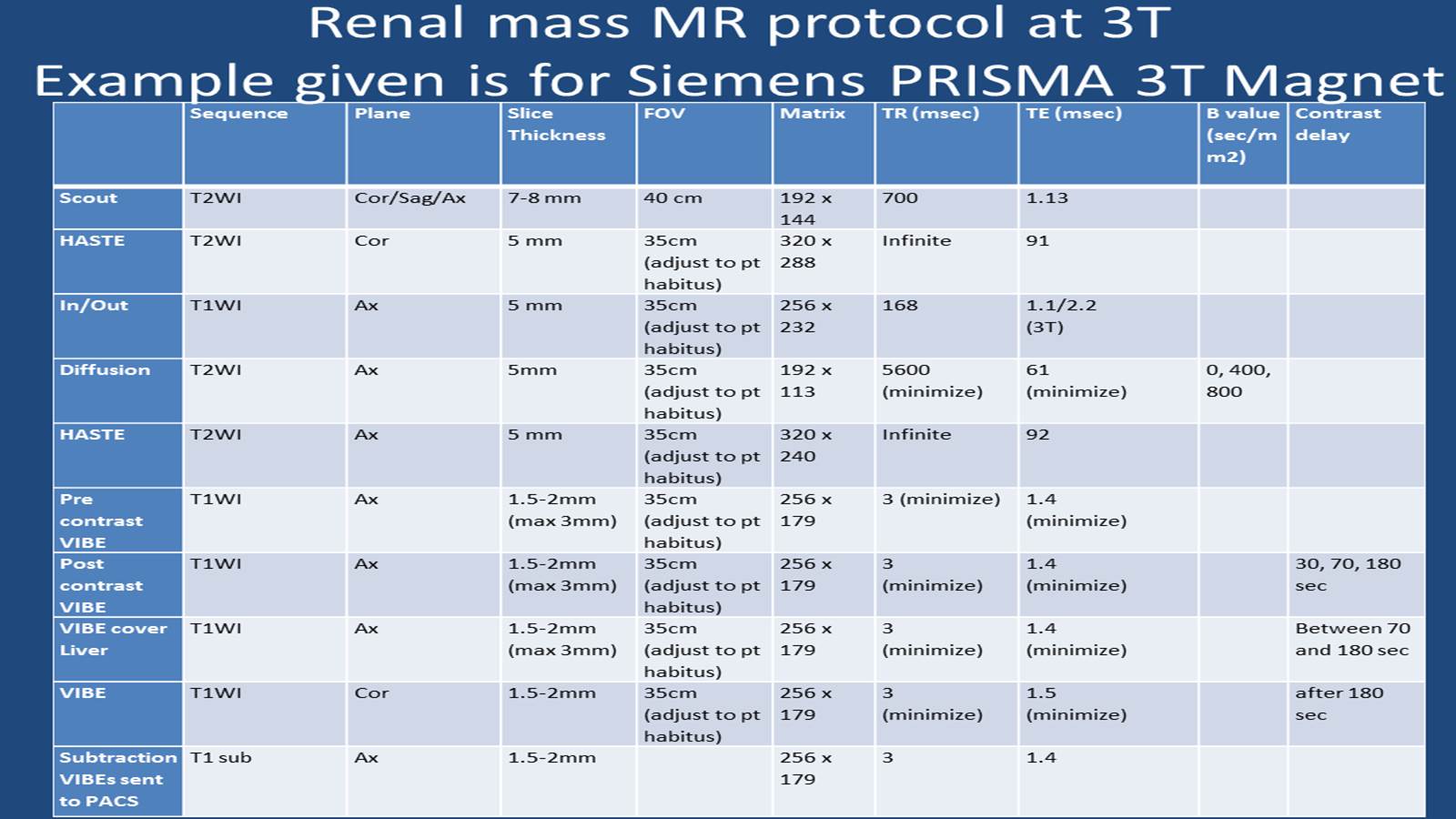
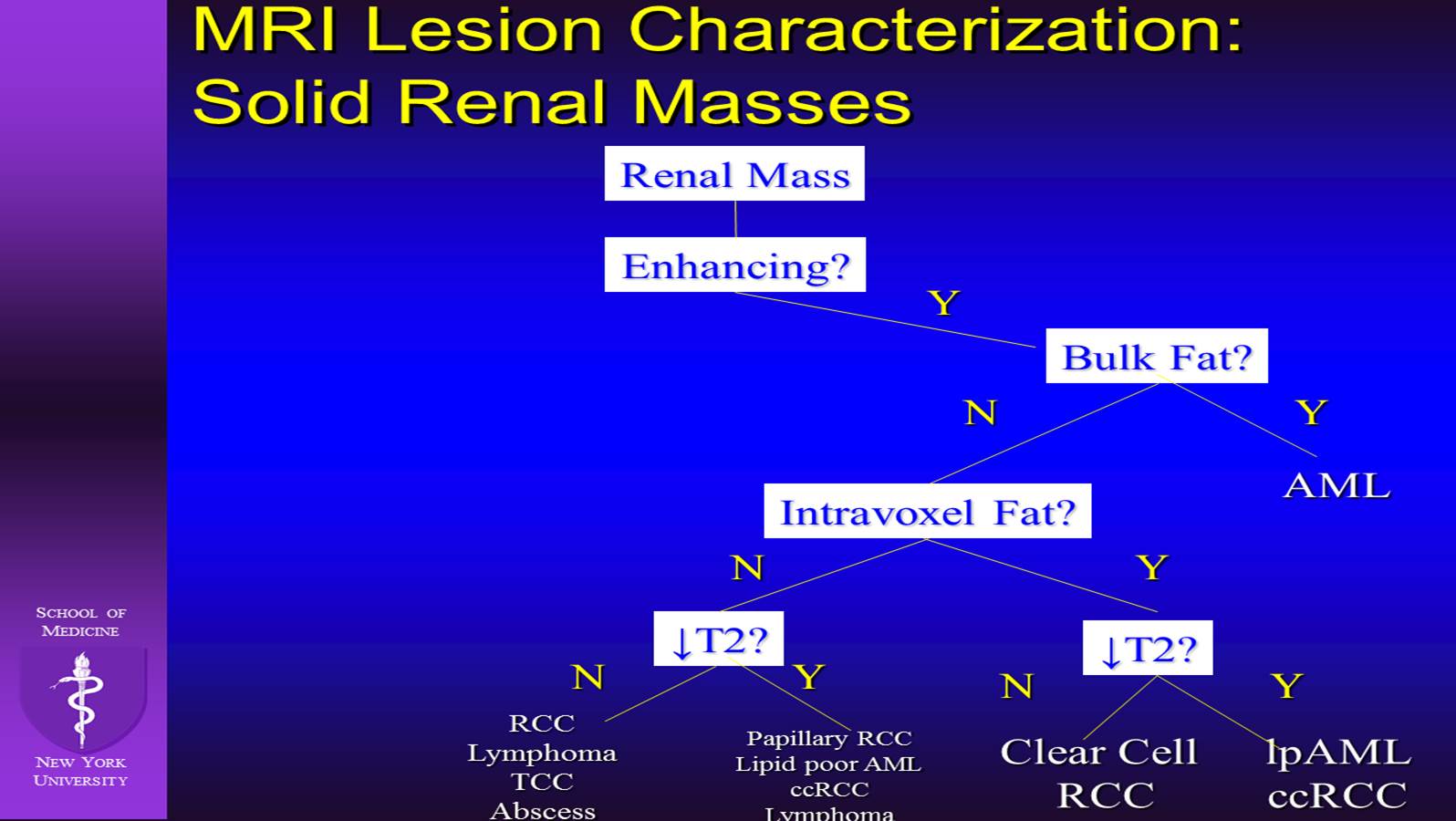
This clinically oriented talk will review the Renal Mass MRI technique/scanning protocols at NYU, review the basics in renal mass lesion subtyping and pitfalls in the characterization of renal lesions and the clinical impact therein.
Acknowledgements
No acknowledgement found.References
1: Yoshida R, Yoshizako T, Hisatoshi A, Mori H, Tamaki Y, Ishikawa N, Kitagaki H.
The additional utility of apparent diffusion coefficient values of clear-cell
renal cell carcinoma for predicting metastasis during clinical staging. Acta
Radiol Open. 2017 Jan 1;6(1):2058460116687174. doi: 10.1177/2058460116687174.
PubMed PMID: 28210496; PubMed Central PMCID: PMC5298554.
2: Potretzke AM, Potretzke TA, Bauman TM, Knight BA, Park AM, Mobley JM,
Figenshau RS, Siegel CL. Computed Tomography and Magnetic Resonance Findings of
Fat-Poor Angiomyolipomas. J Endourol. 2017 Feb;31(2):119-128. doi:
10.1089/end.2016.0219. PubMed PMID: 27897036.
3: Davenport MS, Hu EM, Smith AD, Chandarana H, Hafez K, Palapattu GS, Stuart
Wolf J Jr, Silverman SG; Society of Abdominal Radiology Disease Focused Panel on
Renal Cell Carcinoma.. Reporting standards for the imaging-based diagnosis of
renal masses on CT and MRI: a national survey of academic abdominal radiologists
and urologists. Abdom Radiol (NY). 2016 Nov 22. [Epub ahead of print] PubMed
PMID: 27878338.
4: Maxwell AW, Baird GL, Iannuccilli JD, Mayo-Smith WW, Dupuy DE. Renal Cell
Carcinoma: Comparison of RENAL Nephrometry and PADUA Scores with Maximum Tumor
Diameter for Prediction of Local Recurrence after Thermal Ablation. Radiology.
2016 Nov 22:161225. doi: 10.1148/radiol.2016161225. [Epub ahead of print] PubMed
PMID: 27875105.
5: Kelly EF, Leveillee RJ. Image guided radiofrequency ablation for small renal
masses. Int J Surg. 2016 Dec;36(Pt C):525-532. doi: 10.1016/j.ijsu.2016.11.026.
Review. PubMed PMID: 27847290.
6: Mnatzakanian GN, Shinagare AB, Sahni VA, Hirsch MS, Silverman SG. Early-stage
clear cell tubulopapillary renal cell carcinoma: imaging features and distinction
from clear cell and papillary subtypes. Abdom Radiol (NY). 2016
Nov;41(11):2187-2195. PubMed PMID: 27383741.
7: Chen X, Zhu Q, Li B, Cui W, Zhou H, Duan N, Liu Y, Kundra V, Wang Z. Renal
cell carcinoma associated with Xp11.2 translocation/TFE gene fusion: imaging
findings in 21 patients. Eur Radiol. 2017 Feb;27(2):543-552. doi:
10.1007/s00330-016-4421-4. PubMed PMID: 27255396.
8: Gorin MA, Rowe SP, Allaf ME. Oncocytic Neoplasm on Renal Mass Biopsy: A
Diagnostic Conundrum. Oncology (Williston Park). 2016 May;30(5):426-35. PubMed
PMID: 27188673.
9: Shaaban AM, Rezvani M, Tubay M, Elsayes KM, Woodward PJ, Menias CO.
Fat-containing Retroperitoneal Lesions: Imaging Characteristics, Localization,
and Differential Diagnosis. Radiographics. 2016 May-Jun;36(3):710-34. doi:
10.1148/rg.2016150149. PubMed PMID: 27163589.
10: Nakashima K, Kitagawa Y, Izumi K, Mizokami A, Gabata T, Namiki M. Diagnostic
accuracy of pre-operative imaging findings in presumed clinical T1a renal cell
carcinomas. Oncol Lett. 2016 May;11(5):3189-3193. PubMed PMID: 27123087; PubMed
Central PMCID: PMC4840990.
11: Yokoo T, Clark HR, Pedrosa I, Yuan Q, Dimitrov I, Zhang Y, Lingvay I, Beg MS,
Bobulescu IA. Quantification of renal steatosis in type II diabetes mellitus
using dixon-based MRI. J Magn Reson Imaging. 2016 Nov;44(5):1312-1319. doi:
10.1002/jmri.25252. PubMed PMID: 27007212; PubMed Central PMCID: PMC5035175.
12: Davarpanah AH, Spektor M, Mathur M, Israel GM. Homogeneous T1 Hyperintense
Renal Lesions with Smooth Borders: Is Contrast-enhanced MR Imaging Needed?
Radiology. 2016 Jul;280(1):128-36. doi: 10.1148/radiol.16151240. PubMed PMID:
26919441.
Figures