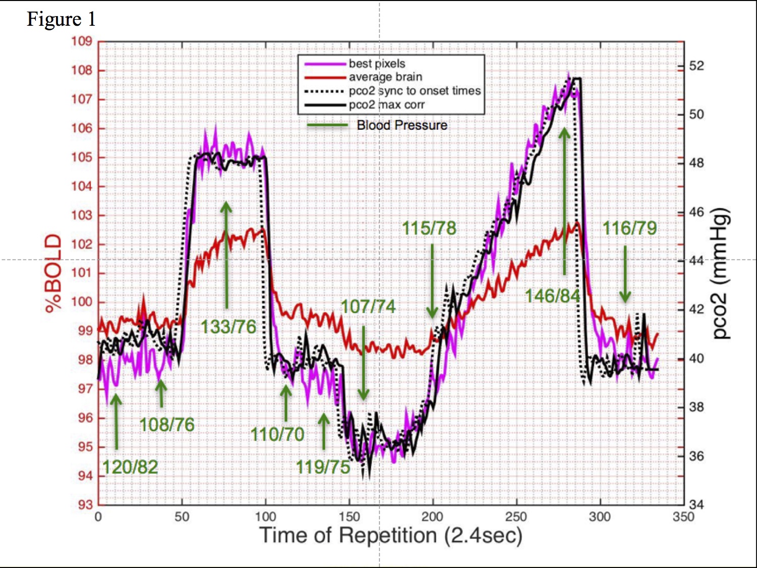
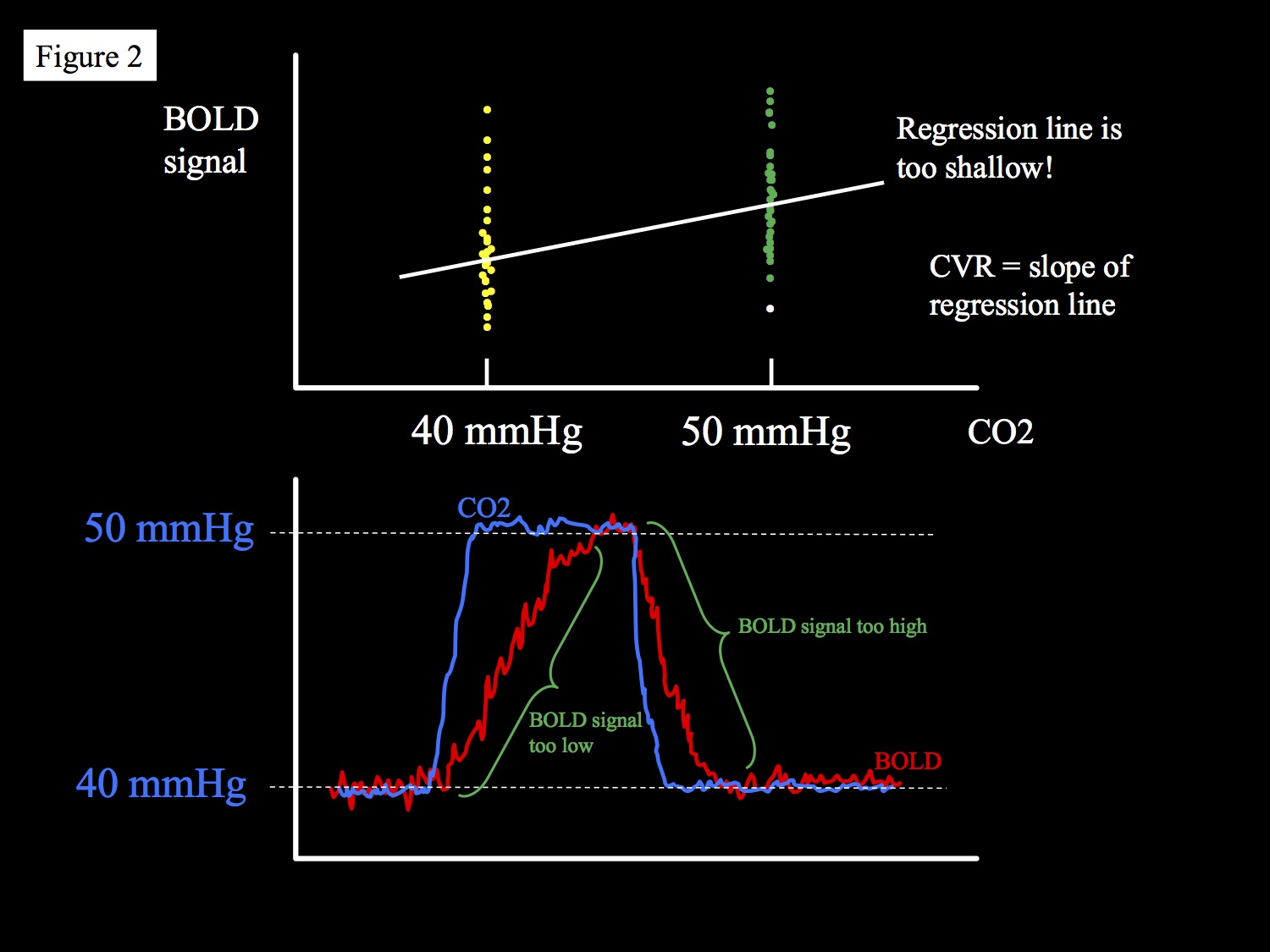
MRI Measurement of Cerebrovascular Reactivity: Clinical Implementation
David Mikulis

Synopsis

The interest in translating MRI mapping of cerebrovascular reactivity (CVR) for the clinical assessment of hemodynamic insufficiency secondary to cerebrovascular disease is increasing. This presentation will focus on the current issues and potential solutions facing widespread dissemination of this methodology. Issues regarding the flow stimulus, flow sensitive pulse sequences, data analysis, and clinically relevant detection thresholds will be presented.

Cerebrovascular reactivity (CVR) is defined as the change in cerebral blood flow per unit change in a vasoactive stimulus. Since the brain depends on a constant supply of oxygen and glucose, and since it is unable to effectively store these energy substrates, uninterrupted delivery is required. In addition to high blood flow requirements during resting conditions, activation of neural networks requires a significant increase in blood flow. Increased blood flow resistance caused by steno-occlusive disease of feeding arteries can result in consumption of vasodilatory reserve that when severe, is associated with an increase in oxygen extraction and an increased risk of major ischemic events as high as six-fold (1). The ability to quantitate CVR in patients with arterial steno-occlusive disease is therefore of interest. Currently, there is no standardized universally accepted method for mapping CVR and there is no consensus on methodology for assessing individual patients. The following is a “wish list” of desirable components for such a protocol: 1. Stimulus a. The stimulus should provide controllable and reproducible changes in blood flow. b. The magnitude and duration of the stimulus should be tolerable and brief. c. Devices used to deliver the stimulus should be easy to implement with minimal set-up time. d. Operation of the device should be as “push-button” as possible enabling widespread application without special training required. 2. Imaging a. The MRI pulse sequence should be able to measure blood flow accurately and quantitatively in the setting of advanced cerebrovascular disease (CVD) with good SNR and no baseline signal drift or be independent of baseline signal drift. b. Spatial resolution should be on the order of cortical thickness or less (2.5 mm). c. Immune to susceptibility effects near the skull base. d. Injection of contrast agents not necessary How much of this protocol can be achieved with current capabilities? 1. Stimulus a. Brief CO2 stimuli 10-15 mmHg above resting levels are safe (2). Elevation of CO2 to these levels generates maps that “appear” to correlate well the underlying vascular pathology but this is a subjective assessment and true CO2 thresholds would require prospective studies with correlation to management outcomes. The need to implement a brief protocol (10 – 12 minutes) and the nature of the existing flow sensitive MR pulse sequences require a CO2 stimulus that can permit averaging similar to that used in task fMRI studies with a minimum of three baselines (resting CO2 conditions) to alleviate the effect of signal baseline drifts on the signal analysis but can also permit full expression of flow changes in slow reacting vascular beds. This requires step changes in the CO2 stimulus similar to fMRI followed by a slower ramp stimulus (figure 1). Note that step changes in CO2 stimuli alone would underestimate CVR values unless the CO2 stimulus was lengthened allowing full evolution of the flow response. This would go against the desired brevity of the protocol. 2. Imaging a. Current pCASL sequences, although desired for the ability to quantitate CBF, are noisy due to the subtracted final images and are not fully delay insensitive to slow flows seen in patients with advanced CVD. b. BOLD is a reasonable surrogate as the flow is near linear in the ranges of CO2 stimuli applied (3). BOLD also has high temporal resolution to measure the speed of vascular responses and easily covers the entire brain. Analysis: 1. Step changes in CO2 enable quantitation and mapping of the speed of the vascular response but can misrepresent true CVR due to long delays in response time (figure 2). 2. Ramp changes in CO2 are more accurate in mapping the true amplitude of the CVR response especially in patients with a slower response times. 3. For clinical assessment of individual subjects, it is important to determine the extent to which the patient’s CVR differs from normal. This can be done by comparing the patient’s CVR maps to an atlas of merged control CVR data. A “Z-map” can then be generated that shows how many standard deviations the patient’s CVR differs from the norm (4). 4. In patients who have follow-up CVR studies, it is important to determine if there are clinically relevant changes in CVR. This is done by creating a new comparison atlas that is made up of controls who have had two CVR studies performed at separate times. A difference image is generated showing spatial differences in CVR between the two time points. This difference takes into account MRI instabilities, subject physiological differences, and differences in the CO2 stimuli (minor if well controlled). The difference maps of each control are then merged into a common brain space creating an interval difference (ID) atlas that maps the mean and standard deviation of the three aforementioned instabilities. A patient’s ID map can then be compared against the control group ID map to define clinically relevant changes in CVR (5). Finally, randomized control studies are needed to substantiate CVR metrics for clinical application including: 1. The magnitude of the stimulus to be applied in order to determine what level is appropriate for detecting clinically relevant hemodynamic insufficiency. Is a 10 mmHg increase in arterial CO2 appropriate? 2. What threshold should be used to determine a clinically relevant CVR deficit? For example, is greater than two standard deviation reductions in CVR the correct threshold for clinical relevance?

Acknowledgements

No acknowledgement found.

References

1. Gupta A, Baradaran H, Schweitzer AD, Kamel H, Pandya A, Delgado D, Wright D, Hurtado-Rua S, Wang Y, Sanelli PC. Oxygen extraction fraction and stroke risk in patients with carotid stenosis or occlusion: a systematic review and meta-analysis. AJNR Am J Neuroradiol. 2014 Feb;35(2):250-5. doi: 10.3174/ajnr.A3668. Review. PubMed PMID: 23945227.

2. Spano VR, Mandell DM, Poublanc J, Sam K, Battisti-Charbonney A, Pucci O, Han JS, Crawley AP, Fisher JA, Mikulis DJ. CO2 blood oxygen level-dependent MR mapping of cerebrovascular reserve in a clinical population: safety, tolerability, and technical feasibility. Radiology. 2013 Feb;266(2):592-8. doi:10.1148/radiol.12112795. PubMed PMID: 23204541.

3. Hoge RD, Atkinson J, Gill B, Crelier GR, Marrett S, Pike GB. Investigation of BOLD signal dependence on cerebral blood flow and oxygen consumption: the deoxyhemoglobin dilution model. Magn Reson Med. 1999 Nov;42(5):849-63. PubMed PMID: 10542343.

4. Sobczyk O, Battisti-Charbonney A, Poublanc J, Crawley AP, Sam K, Fierstra J, Mandell DM, Mikulis DJ, Duffin J, Fisher JA. Assessing cerebrovascular reactivity abnormality by comparison to a reference atlas. J Cereb Blood Flow Metab. 2015 Feb;35(2):213-20. doi: 10.1038/jcbfm.2014.184. Epub 2014 Nov 12. PubMed PMID:25388679.

5. Sobczyk O, Crawley AP, Poublanc J, Sam K, Mandell DM, Mikulis DJ, Duffin J, Fisher JA. Identifying Significant Changes in Cerebrovascular Reactivity to Carbon Dioxide. AJNR Am J Neuroradiol. 2016 May;37(5):818-24. doi: 10.3174/ajnr.A4679. PubMed PMID: 26846924.

Figures

Figure 1 - Step and ramp CO2 stimulus showing end-tidal pCO2 values, the BOLD signal response (best responding voxels and average brain responses), and blood pressure changes during the CO2 stimulus in a patient with moyamoya disease. Blood pressure is monitored as shown. Overall scan time = 13.6 minutes.

Figure 2 - The effect of measuring CVR in vascular beds with delayed vasodilatory responses. BOLD signal is acquired every 2 seconds. 10 mmHg step changes in CO2 occur in 1-2 breaths. In disease states the time to reach maximum BOLD signal may be delayed and return to baseline is delayed. This means that the mean BOLD signal during the maximum stimulus is abnormally low. Similarly, the BOLD signal at rest is artificially increased due to the delay in the return to baseline from hypercapnia. The slope of the regression line which, by definition is CVR, is lower than normal. The effect can also influenced by time shifts of the BOLD signal against the PCO2 regressor used to maximize the correlation coefficient.

Proc. Intl. Soc. Mag. Reson. Med. 25 (2017)