Ultrasound Neuromodulation with MRI for Brain Cirtcuitry in Non-Human Primates
Charles Caskey1, Vandiver Chaplin1, Pai-Feng Yang1, William Grissom1, Tony Phipps1, Allen Newton1, John Gore1, Li Min Chen1, Wolf Zinke2, and Jeffrey Schall2
1Vanderbilt University Institute of Imaging Science, Nashville, TN, United States, 2Psychology, Vanderbilt University, Nashville, TN, United States
Synopsis
In this presentation, I will discuss ongoing work where we are using ultrasound in conjunction with fMRI to modulate and subsequently image brain circuits in non-human primates.
Objectives
Brain stimulation is an effective approach to study neural circuits; however, all stimulation methods to date are either highly invasive (intracortical electrical stimulaton or pharmacological stimulation) or have poor spatial resolution (transcranial magnetic stimulation or direct current stimulation). Focused ultrasound (FUS) has the potential to provide non-invasive neuromodulation with high spatial resolution throughout the brain. In this study, we have developed and applied tools to stimulate the non-human primate cortex while observing changes in behavior and blood oxygen level dependent functional magnetic resonance imaging (BOLD fMRI). We are examining this in two non-human primate models: frontal eye field (FEF) and somatosensory cortex. In this presentation, we provide updates on this ongoing work and discuss practical considerations for integrating ultrasound into the MR environment.Methods: Studies in behaving macaques
A single element focused transducer was used to stimulate FEF, a key node in the eye movement and attention systems, while monkeys performed a visual search task to shift gaze to a target item among distractors. We applied pulsed FUS stimulation through a 2-cm craniotomy for 300 ms prior to the presentation of the search array (Figure 1a; center frequency 500 kHz, total duration 300 ms, repetition frequency 2 kHz, pulse duration 0.25 ms). Saccade response times and intracranial EEG recordings were acquired in two monkeys performing a visual search task while we alternated 10 min blocks with FUS stimulation ON and OFF. With each NHP, we carried out experiments at peak negative pressures of 250 kPa and experiments at 425 kPa.Methods: Combining FUS neuromodulation and fMRI
We have applied ultrasound to the macaque S1/insula region during fMRI using the stereotactic frame developed as part of this project. The system holds the monkey in place in the sphinx position within a human 7T MRI and can accommodate a single element neuromodulation transducer. The transducer is placed under image guidance and the ultrasound beam can be localized by imaging the acoustic radiation force or small temperature rise.Results
In studies of behaving macaques, FUS stimulation at both pressures generated no significant warming of brain tissue (<1.5°C). FUS stimulation at 425 kPa slowed saccade response time (SRT) by 5 ms in a spatially specific manner (Figure 1b, p<0.001). We also found that the higher pressure (425 kPa) elicited a reliable reduction in the magnitude of an event-related potential known as N2pc, which indexes attention; the effect of FUS arose ~90 ms after stimulus onset (Figure 1c). These findings demonstrate the feasibility of FUS modulation of attention and performance in monkeys and suggest potential dose dependence for FUS neuromodulation and spatial selectivity based on the location of stimulation. In FUS-fMRI studies of the macaque somatosensory system, we have detected reproducible tactile stimulus evoked BOLD fMRI activations in expected areas S1 and S2 regions (Figure 2, first two rows). These studies were done in the presence of the inactive FUS stimulation device and validate the ability to detect BOLD fMRI near the transducer and coupling material that can generate artifacts. When applying FUS with the beam in the S2/insula region, fMRI signal changes were detected at the stimulated region as well as in S1 region (Figure 2, last row). No detectable temperature elevation was observed in these studies, and FUS stimulation effects appear to be robust in relevant regions spready across adjacent image slices.Conclusion
Our findings demonstrate the feasibility of FUS modulation of in monkeys and the ability to track these changes using standard behavioral, electrophysiological, and fMRI measurements.Acknowledgements
The authors acknowledge funding from NIH grants R01 MH111877 and R24 MH109105.References
No reference found.Figures
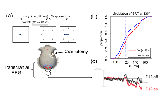
Figure 1. (a) Experimental setup using a single element transducer through a craniotomy during a reward-based task.
During the task, the macaque was fixated on a point in the center of a screen. The primate was required to make an
eye movement towards a presented target item and maintain the gaze for 1 second to receive a juice reward. (b)
Cumulative density function of SRT in one monkey to a target located 135° off center shows robust slowing in saccade
times when FUS is on (p<0.001). (c) In stimulation blocks, FUS was delivered prior target onset, eliciting a modulation
in the mN2PC attention related LFP.
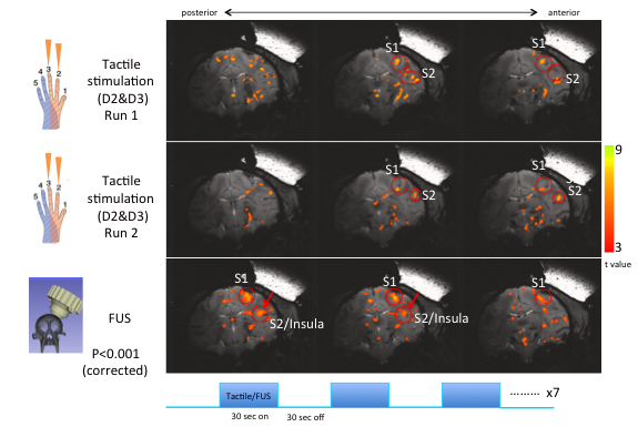
Figure 2. During tactile stimulation of D2 and D3, we have observed robust BOLD fMRI response in S1 and S2 (top
two rows). Upon application of focused ultrasound to the S2/insula region, we see BOLD fMRI changes in the
stimulated region as well as S1 (bottom row). During the experiments, stimulation (either vibrotactile or ultrasound
stimulation at 250 kHz) was applied in 30 second intervals (bottom row).