Beyond Proton MRI: 19F MRI & More
1Radiology, University of Texas Southwestern Medical Center
Synopsis
19F NMR offers exceptional insights for diverse physiological and pharmaceutical investigations. High sensitivity and lack of interfering background signal in the body have enabled the observation of exogenously administered agents and their metabolites. 19F exhibits a large chemical shift range, which is exquisitely sensitive to the microenvironment. In addition to chemical shift, relaxation processes (R1 and R2), and chemical exchange may be tailored to be responsive to a parameter of interest such as pO2, pH, metal ion concentrations, transgene/enzyme activity or hypoxia. I will review 19F NMR/MRI as a foundation for diverse applications and recent innovations.
Highlights
19F NMR exhibits exquisite sensitivity to the microenvironment with opportunities to exploit chemical shift and relaxation parameters
19F NMR exhibits high intrinsic sensitivity and long term molecular stability
19F NMR experiences natural background signal
Notable applications:
i) Quantitative dynamic oximetry
ii) Ions including pH
iii) Enzyme activity
iv) Cell tracking
Background
Fluorine NMR offers many attractive features, most notably detection sensitivity approaching that of protons. 19F has a nuclear spin I=½, a gyromagnetic ratio of 40.05 MHz/T, and is 100% naturally abundant (1). 19F NMR is particularly attractive for in vivo applications since there is essentially no endogenous 19F signal from tissues. Thus, fluorinated reporter molecules or drugs may be introduced into the body and detected readily with high sensitivity and without background interference. Several expansive reviews devoted to 19F NMR exist (2-7) (8), as well as a recent book devoted to the topic (9).
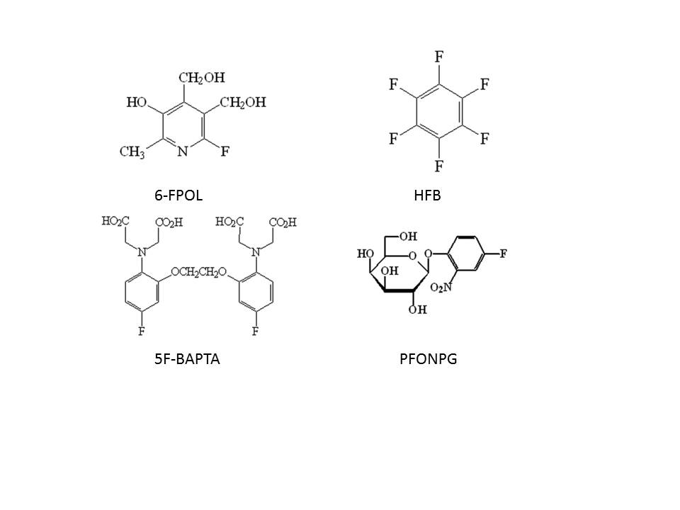
19F is exceptionally sensitive to molecular and microenvironmental changes as exemplified by the many 19F-based reporter molecules designed to interrogate physiological phenomena in vivo including pO2, pH, and [Ca2+] (2, 10-12) (see Figure 1). Fluorine requires millimolar concentrations, as opposed to picomolar typical of the PET tracer 18F (13), but agents labeled with 19F may be traced over hours to days and even weeks allowing assessment of long term pharmacokinetics or evolution of pathophysiology, such as hypoxiation accompanying tumor growth (14, 15).
NMR can provide quantitative measurements based on signal integration. In principle, the larger the number of equivalent 19F atoms the stronger the signal. Trifluoromethyl (-CF3) moieties are popular, since they are metabolically inactive and provide a single non-coupled signal. Six equivalent fluorines are observed for an isopropyl group as exploited in the nitroimidazole hypoxia reporter CCI-103F (16) and tris trifluoromethyl (t-butyl) group provides 9 equivalent fluorines (17, 18). Twelve equivalent fluorines were used by Takaoka et al. (19), who evaluated the relative merits of varying the number of fluorine atoms. Molecular symmetry may also be exploited in molecules such as hexafluorobenzene (HFB) and perfluoro-15-crown-5-ether (15c5), each of which exhibits a single resonance frequency, and has been exploited for in vivo oximetry (20, 21). Ultimately, local 19F concentration is particularly relevant, and thus agents which accumulate at specific targets may be most useful, e.g., perfluorocarbon emulsions tend to accumulate in the reticuloendothelial system (RES) (22). High local concentration has also been achieved by direct injection of reporter molecule into tissue of interest (e.g., hexafluorobenzene in tumors for oximetry (FREDOM) (10, 20)) or pre-labeling of cells with PFC emulsion prior to injection (23).
The 19F atom may modulate molecular properties, most notably hydrophobicity and this becomes more significant for multiple fluorines, as encountered in CF3 groups. In addition, the electronegativity can alter ionization of adjacent groups such as carboxyl, phenol, and amine. Several pharmaceuticals, agrochemicals and pesticides include fluorine and have been examined by 19F NMR (5, 24). It should be noted that CF3 groups tend to resist degradation, but mono and difluoro groups may yield highly reactive degradation products acting as potential enzyme suicide inhibitors (e.g., fluoroacetate is highly toxic).
Applications
i) Oximetry: Perhaps the most sustained application of 19F NMR has been to assess tissue oxygenation. Quantitative 19F MR oximetry has been developed over many years based on the sensitivity of the spin-lattice relaxation rate (R1 =1/T1) of perfluorocarbons (PFCs) to oxygen concentration (25). PFCs exhibit very high gas solubility and high density, while being hydrophobic, essentially inert, and non-toxic. 19F MR oximetry is based on the ideal liquid gas interaction with oxygen giving a linear dependence for the 19F spin lattice relaxation rate, R1 = A + B pO2, as reported and summarized for many different PFCs (10, 26). The relationship remains linear across the whole range of pO2 values including hyperbaric conditions (27). Since PFCs are exceedingly hydrophobic, ions and proteins do not dissolve and therefore calibration curves established in vitro may be used in vivo (28-31).
Oximetry based on PFCs is an effective gold standard for validating other methods, but it must be noted that relaxivity is also sensitive to both field and temperature dependent, and thus, pertinent calibration curves are required (20, 31, 32). The temperature dependence means that even a relatively small error in temperature estimate can introduce a sizable discrepancy into the apparent pO2 for many PFCs. The dependence is however particularly small for HFB, where a 1 oC error in temperature estimate should only cause 0.1 Torr error in pO2 at 4.7 T and 37 oC when the actual pO2 is about 5 Torr (10).
pO2 distributions have been reported using 19F MRI in diverse tumors in rats and mice (33-38) revealing hypoxic fractions ranging from HF10 = 16% in small R3327-H tumors to 83% in large R3327-AT1 tumors on anesthetized rats breathing air (39). The most common studies have involved hyperoxic gas breathing challenges (35, 40) (e.g., Figure 2). Other studies have examined androgen dependence via castration (38), vasoactive agents (41) and vascular disrupting agents (VDAs) (42-44). Two studies are particularly notable: i) Estimates of pO2 and modulation of tumor hypoxia have been shown to be consistent with modified tumor response to irradiation (45, 46). ii) The study of arsenic trioxide by Gallez et al. is potentially paradigm shifting since VDAs are expected to cause vascular shutdown and consequent hypoxia, whereas 19F MRI revealed elevated pO2 at low doses, likely associated with reduced oxygen consumption due to mitochondrial impairment (37).
19F MR oximetry can provide maps of tissue oxygenation and assess dynamic changes in response to interventions. By contrast oxygen electrodes would typically only provide dynamic measurement at a single location, or alternatively provide maps at a single time point. 19F oximetry is best emulated by analogous proton MR oximetry and the concept PISTOL (Proton Imaging of Siloxanes to map Tissue Oxygenation Levels) was developed (47); like 19F oximetry, it can provide dynamic pO2 maps with similar spatial and temporal resolution, but does require water and fat suppression. Currently, proton MR is far more widely available than 19F MR, particularly when considering potential applications to larger animal or man. Both approaches generally use direct injection of reporter molecule in tumors, though many studies have reported 19F oximetry following systemic administration of PFC emulsions.
ii) Detection of ions: metals and pH Diverse ions have been interrogated using 19F NMR reporter molecules. In most cases reporter molecules were designed to undergo a binding-dependent chemical shift. The earliest example of a metal ion reporter was probably 5F-BAPTA (5,5-difluoro-1,2-bis(o-aminophenoxy)ethane-N,N,N',N'-tetraacetic acid), which shows Δδ~6.0 ppm chemical shift upon binding Ca2+ (12). Metal ion binding is often in the slow exchange regime, so that separate signals are seen for the free and metal ion bound moieties, with chemical shift difference of several ppm. 5F-BAPTA has been used to measure [Ca2+] in cell cultures (48), perfused tissue slices (49) and the perfused beating hearts, revealing calcium transients during the myocardial cycle (50, 51).
pH-sensitive 19F NMR indicators were pioneered by Deutsch et al., notably exploiting a series of fluoroalanines to investigate intra- and extracellular pH (52, 53). The relatively small chemical shift range (~ 2 ppm) is quite typical of aliphatic reporter molecules, while aromatic reporter molecules can have a much larger chemical shift response (often approaching 5 to 10 ppm) (54, 55). The vitamin B6 analogue fluoropyridoxol (6-FPOL) readily penetrates red blood cells so that both intra- and extracellular pH (pHi and pHe) could be measured simultaneously in whole blood (56). Measurements were also readily performed in perfused rat hearts (57). However, FPOL does not appear to enter most tumor cells (4).
To enhance SNR, a pH-sensitive CF3 moiety was introduced in place of the F atom, but the chemical shift response of 6-trifluoromethylpyridoxol (CF3POL) was found to be much smaller, as expected since electronic sensing must be transmitted through an additional C-C bond (58). The pKa was better suited to normal tissue physiology and tumor (pKa ~ 6.8 vs. 7.4). CF3POL occurred exclusively in the extracellular compartment (58). While this confounded the ability to directly assess transmembrane pH gradients, it has the potential advantage of defining which compartment is being observed. This can be important since tumors are often heterogeneous with broad resonance peaks and signals representing pHi and pHe may not be resolved. A fluoroaniline sulphonamide (ZK150471) was used to measure tumor pH in mice and rats (59)(60, 61) and it is also restricted to the extracellular compartment. We have generally used sodium trifluoroacetate, as a non-titrating chemical shift reference, but an intramolecular chemical shift reference is feasible (62)(61).
19F NMR has provided unique insights into transmembrane pH gradients in vivo, but poor SNR, generally precludes spatial resolution for assessing tissue heterogeneity.
iii) Proteomics 19F NMR has long been applied to evaluating pharmacology in terms of catabolic and anabolic conversions of drugs and pro-drugs (63-65). Notably, the popular chemotherapeutic 5-FU is converted to multiple products such as 5-fluoronucleotides, 5,6-dihydrofluorouracil, and α-fluoro β-alanine and the relative processes may influence therapeutic efficacy. 19F NMR was also applied to 5-FC to examine expression of cytosine deaminase (CD) in combination with gene therapy (66).
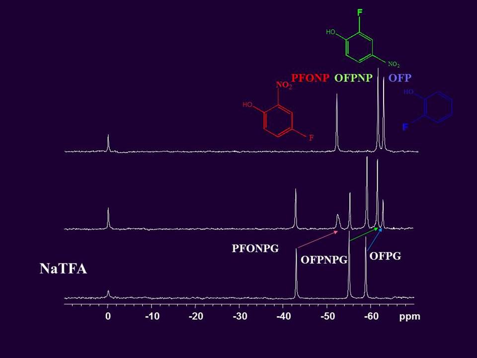
Other specific enzyme activity reporter agents have been demonstrated, notably for β -galactosidase (lacZ). A simple fluorinated analog of the traditional colorimetric yellow reporter ONPG (o-nitrophenylgalactoside) provided an initial 19F NMR active analogue (67). The prototype molecule used a para fluoroaryl substituent in 4-fluoro-2-nitrophenyl β-D-galactopyranoside (PFONPG) (67) providing a single 19F NMR signal with a narrow linewidth and good stability in solution. Enzyme activated cleavage of the glycosidic bond produced a substantial chemical shift Δδ > 3.6 ppm, which was observed in stably transfected human tumor xenografts in mice (67-69). β-gal is reported to exhibit extremely broad substrate specificity (promiscuity) allowing a range of 19F labeled substrates. By varying the orientation of the F atom on the aglycone substrates are produced with unique chemical shifts, also yielding products with unique chemical shift so that multiple agents can be detected simultaneously, potentially facilitating in vivo proteomics (70) (see Figure 3).
iv) Cell tracking Incubation of cells with PFC emulsion can label them and allow tracking in vivo (71) as demonstrated in various cells, tissues and locations (23, 71-77). Since various PFCs can form stable emulsions, different cell populations can be uniquely labeled and observed with spectral selective imaging allowing assay of distributions (Figure 3) (74). This method is being developed primarily for tracking stem cell migration and retention, but also allows oximetry (15). Initially the approach provides optimal signal to noise from the cells and assuredly provides intracellular measurements, though following cell division the signal density of fluorine per cell is reduced and cell death will lead to redistribution potentially to macrophages. The application of MRI for cell tracking will be covered in greater detail in the later educational presentation by Dr. Bulte. The primary advantage of 19F yields over SPIOs is that positive signal is observed, as opposed to susceptibility induced signal voids.
Context
19F NMR offers unique insights into physiology and pharmacology, but does require introduction of exogenously labeled molecules. In some cases, similar information may be obtained based on endogenous materials. While cation-binding diaza-18-crown-6 agents were developed to selectively detect sodium ions, 23Na MRI can be applied directly, though chemical shift agents may be required to differentiate intra- and extracellular signals (78, 79). pH has been measured using 31P NMR of inorganic phosphate (Pi), with the additional benefit that phosphocreatine (PCr) provides a chemical shift reference and other so-called high energy metabolites, notably adenosine triphosphate (ATP) may be detected simultaneously (80-82). 31P NMR signals are often broad and poorly resolved and thus 19F agents have been used in specific situations. Selective signal enhancement over background can be provided by isotopic enrichment, whereby substitution with deuterium can potentially provide 6,400 fold signal gain, though it is subject to kinetic isotope effects (83). 13C is widely applied with minimal kinetic isotope effects, potential 100-fold signal gain and wide chemical shift range so that multiple molecules may be detected simultaneously (84, 85). Moreover, incorporating hyperpolarization may yield as much as 60,000 fold signal enhancement (85, 86) though the signal is transient, decaying rapidly.Opportunities
19F NMR potentially offers unique insights, but its widespread application remains thwarted by SNR. This is an active area of research and promising opportunities are presented as follows. Interleaved and simultaneous detection of 19F and 1H MRI has been developed with dual tuned coils, particularly at low field (87). Dual acquisition should be useful to allow simultaneous detection of dynamic changes, e.g., pharmacokinetics of a fluorinated drug (uptake, clearance, and metabolism) together with pharmacodynamics of its effect (e.g., vascular perturbation). Similar to the goal for PET/MRI (88, 89). NMR visibility is influenced by molecular motion, whereby restricted motion causes line broadening. Several examples have now shown that disruption of aggregate nanostructures produced enhanced 19F NMR signal (19, 90, 91). Judicious incorporation of protein ligands led to recognition driven disassembly of nanoprobes accompanied by strong signal gain. This approach has also been explored to assess pH (92). Three-dimensional compressed sensing 19F MRI methods have been reported to quantify the macrophage burden in a localized wounding-inflammation mouse model in vivo; at 8-fold image acceleration, the 19F signal distribution was reported to be accurately reproduced without pronounced image degradation, with no loss in signal-to-noise ratio. (93)Acknowledgements
Studies in my lab have been supported over the years by NIH (R01 CA139043-01A1; P30 CA142543; EB015908) and CPRIT (RP140285; RP 140399). I am grateful to Dr Donghan Yang for proofreading this review.References
1. Dolbier, W. R. (2009) Guide to Fluorine NMR for Organic Chemists Wiley, Hoboken, NJ
2. Yu, J.-X., Hallac, R. R., Chiguru, S., and Mason, R. P. New frontiers and developing applications in 19F NMR. Progr. Nucl. Magn. Reson. Spectrosc. 2013; 70, 25-49.
3. Kodibagkar, V. D., Hallac, R., Zhao, D., Yu, J.-X., and Mason, R. P. (2012 ) 19F NMR: clinical and molecular imaging applications. Vol. 6 Fluorine in Pharmaceutical and Medicinal Chemistry: From Biophysical Aspects to Clinical Applications pp. 461-524, World Scientific Publishing Company, London 4. Yu, J. X., Kodibagkar, V., Cui, W., and Mason, R. P. 19F: a versatile reporter for non-invasive physiology and pharmacology using magnetic resonance. Curr. Med. Chem. 2005; 12, 818-848.
5. Ruiz-Cabello, J., Barnett, B. P., Bottomley, P. A., and Bulte, J. W. M. Fluorine (19F) MRS and MRI in biomedicine. NMRBiomed. 2011; 24, 114-129.
6. Ahrens, E. T., and Zhong, J. In vivo MRI cell tracking using perfluorocarbon probes and fluorine-19 detection. Nmr in Biomedicine 2013; 26, 860-871. 7. Wickline, S. A., Mason, R. P., Caruthers, S. D., Chen, J., Winter, P. M., Hughes, M. S., and Lanza, G. M. (2010) Fluorocarbon Agents for Quantitative Multimodal Molecular Imaging and Targeted Therapeutics. In Molecular Imaging: Principles and Practice (Weissleder, R., Gambhir, S. S., Ross, B. D., and Rehemtulla, A., eds) pp. 542-573, BC Decker Inc., Hamilton, Ontario, Canada
8. London, R. E. (1994) In vivo NMR studies utilizing fluorinated NMR probes. In NMR in Physiology and Biomedicine (Gillies, R. J., ed) pp. 263-277, Academic, San Diego
9. Flogel, U., and Ahrens, E. (2016) Fluorine Magnetic Resonance Imaging, Pan Stanford
10. Zhao, D., Jiang, L., and Mason, R. P. Measuring Changes in Tumor Oxygenation. Methods Enzymol 2004; 386, 378-418.
11. Taylor, J. S., and Deutsch, C. J. Fluorinated a-methylamino acids as 19F NMR indicators of intracellular pH. Biophys. J. 1983; 43, 261-267.
12. Smith, G. A., Hesketh, R. T., Metcalfe, J. C., Feeney, J., and Morris, P. G. Intracellular Calcium Measurements by F-19 Nmr of Fluorine-Labeled Chelators. Proc. Natl. Acad. Sci. (USA)-Biol. Sci. 1983; 80, 7178-7182.
13. Willmann, J. K., van Bruggen, N., Dinkelborg, L. M., and Gambhir, S. S. Molecular imaging in drug development. Nature Rev. Drug Disc. 2008; 7, 591-607.
14. Mason, R. P., Antich, P. P., Babcock, E. E., Constantinescu, A., Peschke, P., and Hahn, E. W. Non-invasive determination of tumor oxygen tension and local variation with growth. Int. J. Radiat. Oncol. Biol. Phys. 1994; 29, 95-103.
15. Kadayakkara, D. K. K., Janjic, J. M., Pusateri, L. K., Young, W. B., and Ahrens, E. T. In Vivo Observation of Intracellular Oximetry in Perfluorocarbon-Labeled Glioma Cells and Chemotherapeutic Response in the CNS Using Fluorine-19 MRI. Magn. Reson. Med. 2010; 64, 1252-1259.
16. Cline, J. M., Rosner, G. L., Raleigh, J. A., and Thrall, D. E. Quantification of CCI-103F labeling heterogeneity in canine solid tumors. Int. J. Radiat. Oncol. Biol. Phys. 1997; 37, 655-662.
17. Barhate, N. B., Barhate, R. N., Cekan, P., Drobny, G., and Sigurdsson, S. T. A nonafluoro nucleoside as a sensitive F-19 NMR probe of nucleic acid conformation. Org. Lett. 2008; 10, 2745-2747.
18. Zhao, X., Ng, W. Y., Lau, K.-C., Collis, A. E. C., and Horvath, I. T. Generation of (nonafluoro-tert-butoxy)methyl ponytails for enhanced fluorous partition of aromatics and heterocycles. Phys. Chem. Chem. Phys. 2012; 14, 3909-3914.
19. Takaoka, Y., Kiminami, K., Mizusawa, K., Matsuo, K., Narazaki, M., Matsuda, T., and Hamachi, I. Systematic Study of Protein Detection Mechanism of Self-Assembling 19F NMR/MRI Nanoprobes toward Rational Design and Improved Sensitivity. J. Am. Chem. Soc. 2011; 133, 11725-11731. 20. Mason, R. P., Rodbumrung, W., and Antich, P. P. Hexafluorobenzene: a sensitive 19F NMR indicator of tumor oxygenation. NMRBiomed. 1996; 9, 125-134. 21. Dardzinski, B. J., and Sotak, C. H. Rapid tissue oxygen tension mapping using 19F Inversion-recovery Echo-planar imaging of Perfluoro-15-crown-5-ether. Magn. Reson. Med. 1994; 32, 88-97.
22. Mason, R. P., Antich, P. P., Babcock, E. E., Gerberich, J. L., and Nunnally, R. L. Perfluorocarbon imaging in vivo: A 19F MRI study in tumor-bearing mice. Magn. Reson. Imaging 1989; 7, 475-485.
23. Ruiz-Cabello, J., Walczak, P., Kedziorek, D. A., Chacko, V. P., Schmieder, A. H., Wickline, S. A., Lanza, G. M., and Bulte, J. W. M. In Vivo "Hot Spot" MR Imaging of Neural Stem Cells Using Fluorinated Nanoparticles. Magn. Reson. Med. 2008; 60, 1506-1511.
24. Yu, J.-X., Cui, W., Zhao, D., and Mason, R. P. (2008) Non-invasive physiology and pharmacology using 19F magnetic resonance. In Fluorine and Health (Tressaud, A., and Haufe, G., eds) pp. 198-276, Elsevier B.V.
25. Thomas, S. R. (1988) The biomedical applications of Fluorine-19 NMR. In Magnetic Resonance Imaging (Partain, C. L., Price, R. R., Patton, J. A., Kulkarni, M. V., and James, A. E. J., eds) Vol. 2 pp. 1536-1552, W.B. Saunders Co., London 26. Mason, R. P. Non-invasive physiology: 19F NMR of perfluorocarbon. Art. Cells, Blood Sub. & Immob. Biotech. 1994; 22, 1141-1153.
27. Delpuech, J.-J., Hamza, M. A., Serratice, G., and Stébé, M.-J. Fluorocarbons as oxygen carriers. I. An NMR study of oxygen solutions in hexafluorobenzene. J. Chem. Phys. 1979; 70, 2680-2687.
28. Thomas, S. R., Pratt, R. G., Millard, R. W., Samaratunga, R. C., Shiferaw, Y., Clark, L. C., and Hoffmann, R. E. Evaluation of the Influence of the Aqueous-Phase Bioconstituent Environment on the F-19 T1 of Perfluorocarbon Blood Substitute Emulsions. J. Magn. Reson. Imaging 1994; 4, 631-635.
29. Lai, C.-S., Stair, S., Miziorko, H., and Hyde, J. S. Effect of oxygen and the spin label TEMPO-Laurate on 19F and proton relaxation rates of the perfluorochemical blood substitute FC-43 emulsion. J. Magn. Reson. 1984; 57, 447-452.
30. Eidelberg, D., Johnson, G., Barnes, D., Tofts, P. S., Delpy, D., Plummer, D., and McDonald, W. I. 19F NMR imaging of blood oxygenation in the brain. Magn. Reson. Med. 1988; 6, 344-352.
31. Mason, R. P., Shukla, H. P., and Antich, P. P. In vivo oxygen tension and temperature: Simultaneous determination using 19F spectroscopy of perfluorocarbon. Magn. Reson. Med. 1993; 29, 296-302.
32. Shukla, H. P., Mason, R. P., Woessner, D. E., and Antich, P. P. A comparison of three commercial perfluorocarbon emulsions as high field NMR probes of oxygen tension and temperature. J. Magn. Reson. Series B 1995; 106, 131-114. 33. Song, Y., Constantinescu, A., and Mason, R. P. Dynamic breast tumor oximetry: the development of prognostic radiology. Technol. Cancer Res. Treat. 2002; 1, 471-478.
34. Zhao, D., Jiang, L., Hahn, E. W., and Mason, R. P. Comparison of 1H blood oxygen level-dependent (BOLD) and 19F MRI to investigate tumor oxygenation. Magn. Reson. Med. 2009; 62, 357-364.
35. Xia, M., Kodibagkar, V., Liu, H., and Mason, R. P. Tumour oxygen dynamics measured simultaneously by near infrared spectroscopy and 19F magnetic resonance imaging in rats. Phys. Med. Biol. 2006; 51, 45-60.
36. Magat, J., Jordan, B. F., Cron, G. O., and Gallez, B. Noninvasive mapping of spontaneous fluctuations in tumor oxygenation using F-19 MRI. Med. Phys. 2010; 37, 5434-5441.
37. Diepart, C., Karroum, O., Magat, J., Feron, O., Verrax, J., Calderon, P. B., Gregoire, V., Leveque, P., Stockis, J., Dauguet, N., Jordan, B. F., and Gallez, B. Arsenic Trioxide Treatment Decreases the Oxygen Consumption Rate of Tumor Cells and Radiosensitizes Solid Tumors. Cancer Res. 2012; 72, 482-490.
38. McNab, J. A., Yung, A. C., and Kozlowski, P. Tissue oxygen tension measurements in the Shionogi model of prostate cancer using F-19 MRS and MRI. Magn. Reson. Mater Phys. Biol. Med. 2004; 17, 288-295.
39. Mason, R. P., Zhao, D., Pacheco-Torres, J., Cui, W., Kodibagkar, V. D., Gulaka, P. K., Hao, G., Thorpe, P., Hahn, E. W., and Peschke, P. Multimodality imaging of hypoxia in preclinical settings. QJ Nucl. Med. Mol. Imaging 2010; 54, 259-280.
40. Hallac, R. R., Zhou, H., Pidikiti, R., Song, K., Stojadinovic, S., Zhao, D., Solberg, T., Peschke, P., and Mason, R. P. Correlations of noninvasive BOLD and TOLD MRI with pO2 and relevance to tumor radiation response. Magn. Reson. Med. 2014; 71, 1863-1873.
41. Zhao, D., Constantinescu, A., Jiang, L., Hahn, E. W., and Mason, R. P. Prognostic Radiology: quantitative assessment of tumor oxygen dynamics by MRI. Am. J. Clin. Oncol 2001; 24, 462-466.
42. Mason, R. P., Constantinescu, A., Ran, S., and Thorpe, P. E. (2003) Oxygenation in a human tumor xenograft: manipulation through respiratory challenge and anti-body directed infarction. In Oxygen Transport to Tissue XXII. Proceedings of the 27th annual meeting of the International Society on Oxygen Transport to Tissue (Dunn, J. F., and Swartz , H. M., eds) Vol. 530 pp. 197-204, Kluwer Acad. , New York
43. Zhao, D., Jiang, L., Hahn, E. W., and Mason, R. P. Tumor physiological response to combretastatin A4 phosphate assessed by MRI. Int. J. Radiat. Oncol. Biol. Phys 2005; 62, 872-880.
44. Zhou, H., Hallac, R. R., Lopez, R. R., Denney, R., MacDonough, M. T., Li, L., Liu, L., Graves, E. E., Trawick, M. L., Pinney, K. G., and Mason, R. P. Evaluation of tumor ischemia in response to an indole-based vascular disrupting agent using BLI and 19F MRI. Am J Nucl Med Mol Imaging 2015; 5, 143-153. 4
5. Zhao, D., Constantinescu, A., Chang, C.-H., Hahn, E. W., and Mason, R. P. Correlation of Tumor Oxygen Dynamics with Radiation Response of the Dunning Prostate R3327-HI Tumor. Radiat. Res. 2003; 159, 621-631. 46. Bourke, V. A., Zhao, D., Gilio, J., Chang, C.-H., Jiang, L., Hahn, E. W., and Mason, R. P. Correlation of Radiation Response with Tumor Oxygenation in the Dunning Prostate R3327-AT1 Tumor. Int. J. Radiat. Oncol. Biol. Phys. 2007; 67, 1179-1186.
47. Kodibagkar, V. D., Wang, X., Pacheco-Torres, J., Gulaka, P., and Mason, R. P. Proton Imaging of Siloxanes to map Tissue Oxygenation Levels (PISTOL): a tool for quantitative tissue oximetry. NMRBiomed 2008; 21, 899-907.
48. Schanne, F. A., Moskal, J. R., and Gupta, R. K. Effect of lead on intracellular free calcium ion concentration in a presynaptic neuronal model: 19F-NMR study of NG108-15 cells. Brain Res. 1989; 503, 308-311.
49. Badargoffer, R. S., Thatcher, N. M., Morris, P. G., and Bachelard, H. S. Neither Moderate Hypoxia nor Mild Hypoglycemia Alone Causes Any Significant Increase in Cerebral [Ca2+](I) - Only a Combination of the 2 Insults Has This Effect - a P-31 and F-19 NMR-Study. J. Neurochem. 1993; 61, 2207-2214.
50. Marban, E., Kitakaze, M., Chacko, V. P., and Pike, M. M. Ca-2+ Transients in Perfused Hearts Revealed by Gated F-19 Nmr-Spectroscopy. Circ. Res. 1988; 63, 673-678.
51. Kusuoka, H., Backx, P. H., Camilion de Hurtado, M. C., Azan-Backx, M., Marban, E., and Cingolani, H. E. Relative roles of intracellular Ca2+ and pH in shaping myocardial contractile response to acute respiratory alkalosis. Am. J. Physiol. 1993; 265, H1696-1703.
52. Deutsch, C. J., Taylor, J. S., and Wilson, D. F. Regulation of intracellular pH of human peripheral blood lymphocytes as measured by 19F NMR. Proc. Natl. Acad. Sci. (USA) 1982; 79, 7944-7948.
53. Deutsch, C. J., and Taylor, J. S. Intracellular pH measured by 19F NMR. Ann. NY Acad. Sci. 1987; 508, 33-47.
54. Deutsch, C. J., and Taylor, J. S. New class of 19F pH indicators: fluoroanilines. Biophys. J. 1989; 55, 799-804.
55. Mason, R. P. Transmembrane pH gradients in vivo: measurements using fluorinated vitamin B6 derivatives. Curr. Med. Chem. 1999; 6, 481-499.
56. Mehta, V. D., Kulkarni, P. V., Mason, R. P., Constantinescu, A., Aravind, S., Goomer, N., and Antich, P. P. 6-Fluoropyridoxol: a novel probe of cellular pH using 19F NMR spectroscopy. FEBS Letters 1994; 349, 234-238.
57. Hunjan, S., Mason, R. P., Mehta, V. D., Kulkarni, P. V., Aravind, S., Arora, V., and Antich, P. P. Simultaneous intracellular and extracellular pH measurement in the heart by F-19 NMR of 6-fluoropyridoxol. Magn. Reson. Med. 1998; 39, 551-556.
58. Yu, J. X., Cui, W., Bourke, V. A., and Mason, R. P. 6-Trifluoromethyl Pyridoxine: Novel 19F-NMR pH Indicator for In Vivo Detection. J. Med. Chem. 2012 55, 6814−6821.
59. Aoki, Y., Akagi, K., Tanaka, Y., Kawai, J., and Takahashi, M. Measurement of intratumor pH by pH indicator used in 19F MR spectroscopy. Invest. Radiol. 1996; 31, 680-689.
60. Ojugo, A. S., McSheehy, P. M., McIntyre, D. J., McCoy, C., Stubbs, M., Leach, M. O., Judson, I. R., and Griffiths, J. R. Measurement of the extracellular pH of solid tumours in mice by magnetic resonance spectroscopy: a comparison of exogenous 19F and 31P probes. NMRBiomed. 1999; 12, 495-504.
61. Frenzel, T., Koszler, S., Bauer, H., Niedballa, U., and Weinmann, H. J. Noninvasive in vivo pH measurement using a fluorinated pH probe and fluorine-19 magnetic resonance spectroscopy. Invest. Radiol. 1994; 29, S220-222.
62. He, S., Mason, R. P., Hunjan, S., Mehta, V. D., Arora, V., Katipally, R., Kulkarni, P. V., and Antich, P. P. Development of Novel 19F NMR pH Indicators: Synthesis and Evaluation of a Series of Fluorinated Vitamin B6 Analogs. Bioorg. Med. Chem. 1998; 6, 1631-1639. 63. van Laarhoven, H. W. M., Punt, C. J. A., Kamm, Y. J. L., and Heerschap, A. Monitoring fluoropyrimidine metabolism in solid tumors with in vivo 19F magnetic resonance spectroscopy. Crit. Rev. Oncol. Hematol. 2005; 56, 321-343.
64. Wolf, W., Albright, M. J., Silver, M. S., Weber, H., Reichardt, U., and Sauer, R. Fluorine-19 NMR spectroscopic studies of the metabolism of 5-fluorouracil in the liver of patients undergoing chemotherapy. Magn. Reson. Imaging 1987; 5, 165-169.
65. Reid, D. G., and Murphy, P. S. Fluorine magnetic resonance in vivo: A powerful tool in the study of drug distribution and metabolism. Drug Discov. Today 2008; 13, 473-480.
66. Stegman, L. D., Rehemtulla, A., Beattie, B., Kievit, E., Lawrence, T. S., Blasberg, R. G., Tjuvajev, J. G., and Ross, B. D. Noninvasive quantitation of cytosine deaminase transgene expression in human tumor xenografts with in vivo magnetic resonance spectroscopy. Proc. Natl. Acad. Sci. (USA) 1999; 96, 9821-9826.
67. Cui, W., Otten, P., Li, Y., Koeneman, K., Yu, J., and Mason, R. P. A novel NMR approach to assessing gene transfection: 4-fluoro-2-nitrophenyl-b-D-galactopyranoside as a prototype reporter molecule for b-galactosidase. Magn. Reson. Med. 2004; 51, 616-620.
68. Liu, L., Kodibagkar, V. D., Yu, J.-X., and Mason, R. P. 19F-NMR detection of lacZ gene expression via the enzymic hydrolysis of 2-fluoro-4-nitrophenyl b-D-galactopyranoside in vivo in PC3 prostate tumor xenografts in the mouse. FASEB J. 2007; 21, 2014-2019. 69. Yu, J. X., Kodibagkar, V. D., Liu, L., and Mason, R. P. A 19F NMR Approach using Reporter Molecule Pairs to Assess b-Galactosidase in Human Xenograft Tumors in Vivo. NMRBiomed. 2008; 21, 704 -712.
70. Yu, J. X., Otten, P., Ma, Z., Cui, W., Liu, L., and Mason, R. P. Novel NMR Platform for Detecting Gene Transfection: Synthesis and Evaluation of Fluorinated Phenyl b-D-Galactosides with Potential Application for Assessing LacZ Gene Expression. Bioconj. Chem. 2004; 15, 1334-1341.
71. Ahrens, E. T., Flores, R., Xu, H. Y., and Morel, P. A. In vivo imaging platform for tracking immunotherapeutic cells. Nature Biotechnology 2005; 23, 983-987.
72. Srinivas, M., Turner, M. S., Janjic, J. M., Morel, P. A., Laidlaw, D. H., and Ahrens, E. T. In Vivo Cytometry of Antigen-Specific T Cells Using F-19 MRI. Magn. Reson. Med. 2009; 62, 747-753.
73. Srinivas, M., Morel, P. A., Ernst, L. A., Laidlaw, D. H., and Ahrens, E. T. Fluorine-19 MRI for visualization and quantification of cell migration in a diabetes model. Magn. Reson. Med. 2007; 58, 725-734.
74. Partlow, K. C., Chen, J. J., Brant, J. A., Neubauer, A. M., Meyerrose, T. E., Creer, M. H., Nolta, J. A., Caruthers, S. D., Lanza, G. M., and Wickline, S. A. F-19 magnetic resonance imaging for stem/progenitor cell tracking with multiple unique perfluorocarbon nanobeacons. FASEB J. 2007; 21, 1647-1654.
75. Srinivas, M., Heerschap, A., Ahrens, E. T., Figdor, C. G., and de Vries, I. J. M. F-19 MRI for quantitative in vivo cell tracking. Trends Biotechnol. 28, 363-370.
76. Boehm-Sturm, P., Mengler, L., Wecker, S., Hoehn, M., and Kallur, T. In Vivo Tracking of Human Neural Stem Cells with F-19 Magnetic Resonance Imaging. PLoS One 2011; 6, 77. Bible, E., Dell'Acqua, F., Solanky, B., Balducci, A., Crapo, P. M., Badylak, S. F., Ahrens, E. T., and Modo, M. Non-invasive imaging of transplanted human neural stem cells and ECM scaffold remodeling in the stroke-damaged rat brain by F-19- and diffusion-MRI. Biomaterials 33, 2858-2871.
78. Gupta, R. K. (1987) 23Na NMR spectroscopy of intact cells and tissues. In NMR spectroscopy of cells and organisms Vol. 2 pp. 2-32, CRC Press, Boca Raton
79. Bansal, N., Germann, M. J., Seshan, V., Shires III, G. T., Malloy, C. R., and Sherry, A. D. Thulium1,4,7,10-tetraazacyclododecane-1,4,7,10-tetrakis (methylene phosphate) as a 23Na shift reagent for the in vivo rat liver. Biochemistry 1993; 32, 5638-5645. 80. Stubbs, M., Bhujwalla, Z. M., Tozer, G. M., Rodrigues, L. M., Maxwell, R. J., Morgan, R., Howe, F. A., and Griffiths, J. R. An assessment of 31P MRS as a method of measuring pH in rat tumours. NMR Biomed. 1992; 5, 351-359.
81. Evanochko, W. T., Ng, T. C., and Glickson, J. D. Application of in vivo NMR spectroscopy to cancer. Magn. Reson. Med. 1984; 1, 508-534.
82. Kemp, G. J., Meyerspeer, M., and Moser, E. Absolute quantification of phosphorus metabolite concentrations in human muscle in vivo by 31P MRS: a quantitative review. NMRBiomed. 2007; 20, 555-565.
83. Mason, R. P., and Sanders, J. K. M. In vivo enzymology: a deuterium NMR study of formaldehyde dismutase in Pseudomonas putida F61a and Staphylococcus aureus. Biochemistry 1989; 28, 2160-2168.
84. Mason, R. P., Sanders, J. K. M., Crawford , A., and Hunter, B. K. Formaldehyde metabolism by E. Coli: detection using in vivo 13C NMR spectroscopy of S-(hydroxymethyl) glutathione as a transient intracellular intermediate. Biochemistry, 1986; 25, 4504-4507.
85. Malloy, C. R., Merritt, M. E., and Sherry, A. D. Could C-13 MRI assist clinical decision-making for patients with heart disease? NMRBiomed. 2011; 24, 973-979. 86. Kurhanewicz, J., Vigneron, D. B., Brindle, K., Chekmenev, E. Y., Comment, A., Cunningham, C. H., DeBerardinis, R. J., Green, G. G., Leach, M. O., Rajan, S. S., Rizi, R. R., Ross, B. D., Warren, W. S., and Malloy, C. R. Analysis of Cancer Metabolism by Imaging Hyperpolarized Nuclei: Prospects for Translation to Clinical Research. Neoplasia 13, 81-97.
87. Keupp, J., Rahmer, J., Grasslin, I., Mazurkewitz, P. C., Schaeffter, T., Lanza, G. M., Wickline, S. A., and Caruthers, S. D. Simultaneous Dual-Nuclei Imaging for Motion Corrected Detection and Quantification of 19F Imaging Agents. Magn. Reson. Med. 2011; 66, 1116-1122.
88. Antoch, G., and Bockisch, A. Combined PET/MRI: a new dimension in whole-body oncology imaging? Eur. J. Nucl. Med. Mol. Imaging 2009; 36, 113-120.
89. Buscher, K., Judenhofer, M. S., Kuhlmann, M. T., Hermann, S., Wehrl, H. F., Schafers, K. P., Schafers, M., Pichler, B. J., and Stegger, L. Isochronous Assessment of Cardiac Metabolism and Function in Mice Using Hybrid PET/MRI. J. Nucl. Med. 2010; 51, 1277-1284.
90. Tanaka, K., Kitamura, N., and Chujo, Y. Bimodal Quantitative Monitoring for Enzymatic Activity with Simultaneous Signal Increases in 19F NMR and Fluorescence Using Silica Nanoparticle-Based Molecular Probes. Bioconj. Chem. 2011; 22, 1484-1490. 91. Wang, H., Raghupathi, K. R., Zhuang, J. M., and Thayumanavan, S. Activatable Dendritic F-19 Probes for Enzyme Detection. ACS Macro Lett. 2015; 4, 422-425.
92. Huang, X. N., Huang, G., Zhang, S. R., Sagiyama, K., Togao, O., Ma, X. P., Wang, Y. G., Li, Y., Soesbe, T. C., Sumer, B. D., Takahashi, M., Sherry, A. D., and Gao, J. M. Multi-Chromatic pH-Activatable F-19-MRI Nanoprobes with Binary ON/OFF pH Transitions and Chemical-Shift Barcodes. Angewandte Chemie-International Edition 2013; 52, 8074-8078.
93. Zhong, J., Mills, P. H., Hitchens, T. K., and Ahrens, E. T. Accelerated fluorine-19 MRI cell tracking using compressed sensing. Magn. Reson. Med. 2012; n/a-n/a.
94. Metcalfe, J. C., Hesketh, T. R., and Smith, G. A. Free cytosolic Ca2+ measurements with fluorine labelled indicators using 19F NMR. Cell Calcium 1985; 6, 183-195.
95. Kodibagkar, V. D., Yu, J., Liu, L., Hetherington, H. P., and Mason, R. P. Imaging b-galactosidase activity using 19F chemical shift imaging of LacZ gene-reporter molecule 2-fluoro-4-nitrophenol-b-D-galactopyranoside. Magn. Reson. Imaging 2006; 24, 959-962.
Figures