Case Study: FOCUS
1Electrical & Electronics Engineering, Bilkent University, Ankara, Turkey, 2National Magnetic Resonance Research Center (UMRAM), Bilkent University, Ankara, Turkey, 3Neuroscience Program, Sabuncu Brain Research Center, Bilkent University, Ankara, Turkey
Synopsis
FOCUS is the product name for the technique that achieves reduced FOV imaging using a 2D spatially selective RF excitation. This technique provides increased image resolution while significantly reducing off-resonance induced artifacts for single-shot echo planar imaging (ssEPI) in diffusion weighted imaging (DWI). This presentation will cover the technical details and the product development stages of FOCUS.
Purpose
To provide technical and product development stages for FOCUS, a reduced field-of-view (rFOV) imaging technique that has been used widely and successfully in the clinical settings for diffusion weighted imaging (DWI).Introduction
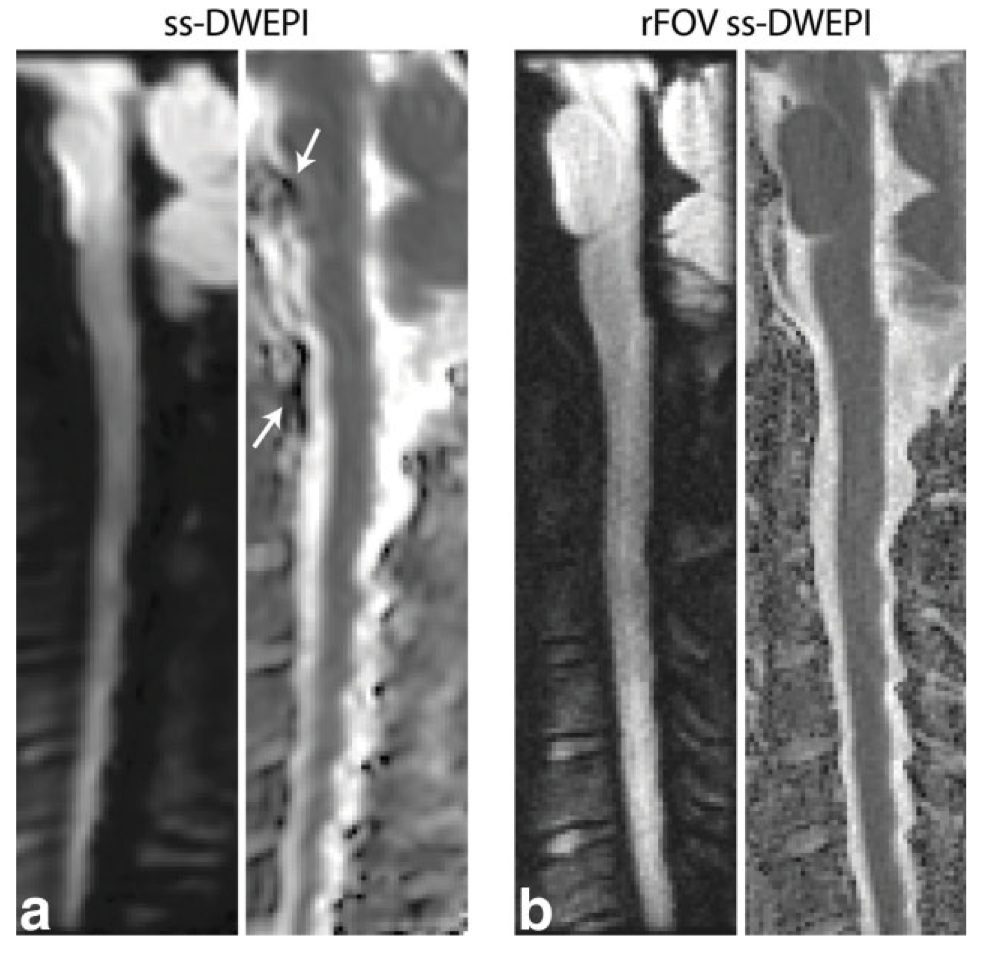
Single-shot echo planar imaging (ssEPI) is currently the most commonly used technique for diffusion weighted imaging (DWI), owing to both its speed and robustness against motion-induced phase errors. The long readout duration in ssEPI, however, makes the images sensitive to off-resonance effects. Multi-shot imaging techniques could alleviate this problem via shortening the echo train, but these techniques are in turn sensitive to motion-induced phase errors that differ from shot to shot in DWI [1]. An alternative solution is reduced field-of-view (rFOV) imaging, which allows imaging only the region of interest without causing aliasing artifacts. Imaging only a small FOV in the phase-encode direction allows a shortening of the echo train in ssEPI, which reduces off-resonance sensitivity and significantly improves image quality. Up to date, rFOV DWI has been proposed via different approach, which include zonal oblique multislice (ZOOM) EPI [2], outer volume suppression [3], parallel imaging with outer volume suppression [4], and 2D selective excitation [5,6].
FOCUS is the product name for the technique that achieves rFOV using a 2D spatially selective RF excitation that provides selectivity in both the phase-encoding and slice directions [5]. We have demonstrated the effectiveness and success of FOCUS DWI over full FOV imaging in various anatomical sites such as the spine [7-8], breast [9-10], prostate [11], and even for fine structures such as nerve roots [12].
This presentation will cover the technical details and the product development stages of FOCUS. The key enabling factors were robustness and ease of use in the clinical routine. Particularly, the collaborative work of the initial developer team, the vendor partner and the clinical team at every stage of the evaluation process will be emphasized.
Acknowledgements
This work was a collaborative work with General Electric Healthcare.References
[1] Anderson A, Gore J. Analysis and correction of motion artifacts in diffusion weighted imaging. Magn Reson Med 1994;32:379–387.
[2] Wheeler-Kingshott CA, Parker GJ, Symms MR, Hickman SJ, Tofts PS, Miller DH, Barker GJ. ADC mapping of the human optic nerve: increased resolution, coverage, and reliability with CSF-suppressed ZOOM-EPI. Magn Reson Med 2002;47:24–31.
[3] Wilm BJ, Svensson J, Henning A, Pruessmann KP, Boesiger P, Kollias SS. Reduced field-of-view MRI using outer volume suppression for spinal cord diffusion imaging. Magn Reson Med 2007;57:625–630.
[4] Heidemann RM, Anwander A, Feiweier T, Kno€sche TR, Turner R. Turner. k-space and q-space: combining ultra-high spatial and angu- lar resolution in diffusion imaging using ZOOPPA at 7 T. Neuro- image 2012;60:967–978.
[5] Saritas EU, Cunningham CH, Lee JH, Han ET, Nishimura DG. DWI of the spinal cord with reduced FOV single-shot EPI. Magn Reson Med 2008;60:468–473.
[6] Finsterbusch J. High-resolution diffusion tensor imaging with inner field-of-view EPI. J Magn Reson Imaging 2009;29:987–993.
[7] Zaharchuk G, Saritas EU, Andre JB, Chin CT, Rosenberg J, Brosnan TJ, Shankaranarayan A, Nishimura DG, Fischbein NJ. Reduced field- of-view diffusion imaging of the human spinal cord: comparison with conventional single-shot echo-planar imaging. AJNR Am J Neu- roradiol 2011;32:813–820.
[8] Andre JB, Zaharchuk G, Saritas E, Komakula S, Shankaranarayan A, Banerjee S, Rosenberg J, Nishimura DG, Fischbein NJ. Clinical evalu- ation of reduced field-of-view diffusion-weighted imaging of the cer- vical and thoracic spine and spinal cord. AJNR Am J Neuroradiol 2012;33:1860–1866.
[9] Singer L, Wilmes LJ, Saritas EU, Shankaranarayanan A, Proctor E, Wisner DJ, Chang B, Joe BN, Nishimura DG, Hylton NM. High-resolu- tion diffusion-weighted magnetic resonance imaging in patients with locally advanced breast cancer. Acad Radiol 2012;19:526–534.
[10] Wilmes LJ, McLaughlin RL, Newitt DC, et al. High-resolution diffusion- weighted imaging for monitoring breast cancer treatment response. Acad Radiol 2013;20:581–589.
[11] Korn N, Kurhanewicz J, Banerjee S, Starobinets O, Saritas E, Noworolski S. Reduced-FOV excitation decreases susceptibility artifact in diffusion-weighted MRI with endorectal coil for prostate can- cer detection. Magn Reson Imaging 2015;33:56–62.
[12] Karampinos DC, Melkus G, Shepherd TM, Banerjee S, Saritas EU, Shankaranarayanan A, Hess CP, Link TM, Dillon WP, Majumdar S. Diffusion tensor imaging and T2 relaxometry of bilateral lumbar nerve roots: feasibility of in-plane imaging. NMR Biomed 2013;26: 630–637.
Figures