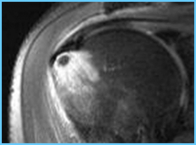
Crystal Deposition Disease
Marcelo Abreu1

1Hospital Mae de Deus

Synopsis

Several 
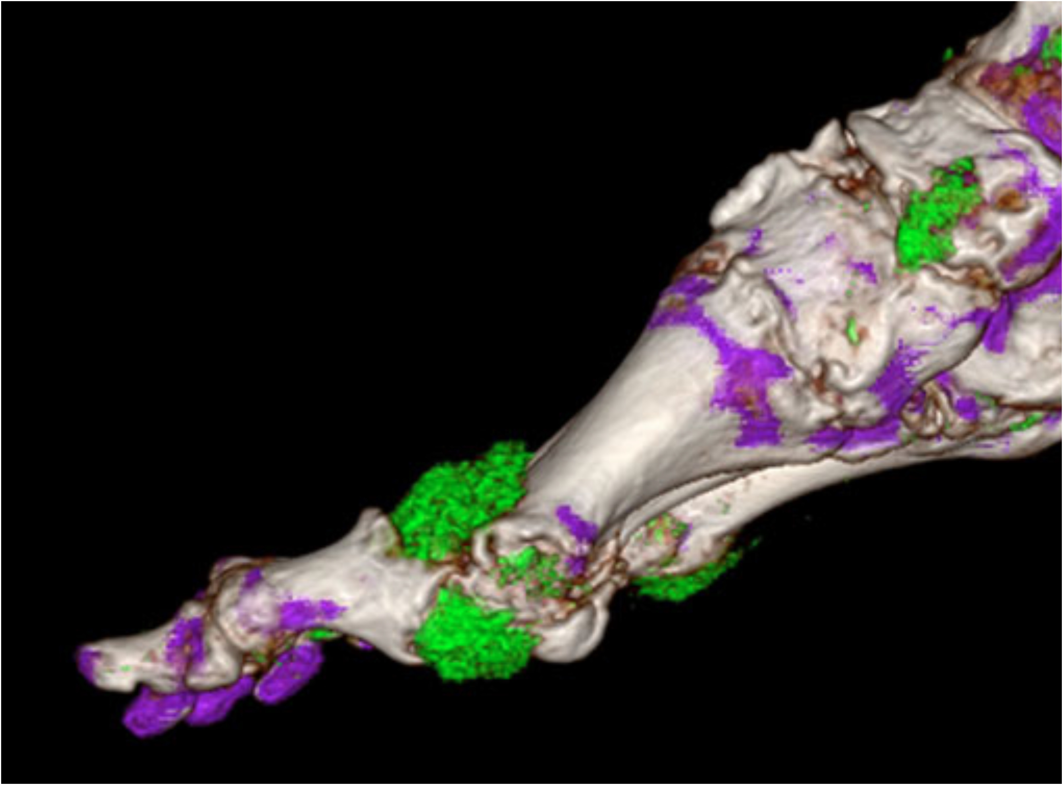
crystals (urate, CPPD and hydroxyapatite) can deposit in tissues of the musculoskeletal system in asymptomatic individuals. I in some cases, those deposits can be associated with diseases such as Gout, Pseudogout and Calcific Tendinitis. MRI examination can be very useful in the diagnosis of such entities, differential diagnosis and help treatment decisions.

Several crystals can deposit in tissues of the muscle skeletal system in asymptomatic individuals, and in some cases, be associated with diseases. There are various imaging features for common crystal-related deposition entities including monosodium urate, calcium pyrophosphate dihydrate as well as hydroxyapatite. Magnetic resonance imaging (MRI) examination features associated with crystal deposition diseases include: a) location (hyaline cartilage and fibrocartilage, tendons, ligaments, bone, synovial membrane), b) morphology (size, soft-tissue signal characteristics), c) surrounding tissue signal alteration, d) arthritic changes (bone, synovial membrane), leading to a deferential diagnosis among those crystal-related deposition entities and other diseases. Monosodium urate monohydrate crystals or uric acid deposits into tissues can cause a disease named Gout. It happens when hyperurecimia is above 6 to 7 mg/dL. Gout is the most common inflammatory arthritis in men over the age of 30 years. Radiographic findings of gout do not occur until the disease has been present for at least 6 or more years. In acute phase MRI findings of the joint effusion and synovial thickening are nonspecific. Gouty tophi are mostly of low- or intermediate-signal intensity on T1W images with variable gadolinium enhancement. The imaging findings of tophi on T2W images are variable. Calcification, fibrosis, or hemosiderin can occur within the tophi. With dual source computed tomography, the uric acid deposits can be differentiated with high accuracy and high imaging contrast. Calcium pyrophosphate dihydrate (CPPD) crystal deposit can be found with a high frequency in older people in the hyaline cartilage, fibrocartilage, tendons and ligaments. Calcification in the spinal ligaments specifically around the odontoid process is common in this entity. Focal calcifications forming conglomerates around joints like tophaceous gout may be also seen. When CPPD crystal deposits become symptomatic it receives the name of Pseudogout, most commonly occurring in knee and wrist. It is often seen in women >80 years old (``disease of octogenarians´´). The shoulder is a non-weight-bearing joint and degenerative changes and narrowing and loss of articular cartilage seen in this joint with the absence of prior injury raises the possibility of CPPD. CPPD deposit may cause arthritis with severe inflammation or may present as destructive joint disease, which may mimic neuropathic joint. The appearance of scapholunate advanced collapse (SLAC) in the wrist is an example of destructive features of pseudogout. Intervertebral disk CPPD crystal deposits can be found in asymptomatic individual but in some more rare cases can cause destructive disc disease. Calcium hydroxyapatite (HA) deposits also can be found in asymptomatic individuals, are often periarticular or intratendinous. Although CPPD is the most common cause of radiographic signs in chondrocalcinosis, both CPPD and calcium hydroxyapatite can be present. On MR imaging, HA deposits are of low signal. Moreover, there is often overlying inflammation and edema, which the radiologist may misinterpret as tenosynovitis or joint synovitis with infection or injury. At high calcium concentrations (above 30% to 40%) susceptibility effects and decreases in proton density dominate, leading to signal intensity loss. However, T1 shortening effects resulting in hyperintensity on T1-weighted images are also present. They have been attributed to surface interaction of protons with calcified tissue. At lower concentrations of calcium, T1 shortening effects dominate, resulting in isointensity or even hyperintensity. Gradient echo sequences best show these calcific foci. The deposition of HA may mimic an acute inflammation. For those patients that are symptomatic, trauma or acutely increased use of the joint seem to be initiating factors. Calcific tendinosis is most commonly seen in the shoulder in the distal supraspinatus tendon insertion at the greater tuberosity of the humerus. Less common locations may include tendons of infraspinatus, subscapularis, and deltoid, wrist, elbow, gluteus maximus, knee, and neck. Rarely erosion of bone adjacent to calcification at the insertion site of the tendons can be visualized.

Acknowledgements

No acknowledgement found.

References

Pascual E, Jovani V. Synovial fluid analysis. Best Pract Res Clin Rheumatol. 2005;19:371-386.

2. Bond JR, Sim FH, Sundaram M. Radiologic case study: Gouty tophus involving the distal quadriceps tendon. Orthopedics. 2004;27:90-92.

3. Miller LJ, Pruett SW, Losada R, et al. Tophaceous gout of the lumbar spine: MR findings. J Computer Assisted Tomography. 1996;20:1004-1005.

4. Chen CKH, Yeh LR, Pan HB, et al. Intra articular gouty tophi of the knee: CT and MRI imaging in 12 patients. Skeletal Radiology. 1999;28:75-80.

5. Yu JS, Chung C, Recht M, et al. MR imaging of the tophaceous gout. AJR Am J Roengtgenol. 1997;168:523-527.

6. McCarty DJ, Hollander JL. Identification of urate crystals in gouty synovial fluid. Ann Intern Med. 1961; 54:452-460.

7. McCarty DJ, Kohn NN, Faires JS. The significance of calcium pyrophosphate crystals in synovial fluid of arthritis patients: the “pseudogout” syndrome. Ann Intern Med. 1962;56:711-737.

8. Zitnan D, Sitaj S. Chondrocalcinosis polyarticularis (familiaris): Roentgenological and clinical analysis.Cesk Rentgenol. 1960;14:27-34.

9. Timms AE, Zhang Y, Russell RG, Brown MA. Genetic studies of disorders of calcium crystal deposition. Rheumatology. 2002;41:725-729.

10. Scotchford CA, Greenwald S, Ali SY. Calcium phosphate crystal distribution in the superficial zone of human femoral head articular cartilage. J Anat. 1992;181:293-300.

11. Doherty M, Dieppe P, Watt I. Low incidence of calcium pyrophosphate dihydrate crystal deposition in rheumatoid arthritis, with modification of radiographic featuers in coexistent disease. Arthritis Rheum. 1984; 27:1002-1009.

12. Steinbach LS, Resnick D. Calcium pyrophosphate dihydrate crystal deposition disease revisited. Radiology. 1996;200:1-9.

13. Giachelli CM. Inducers and inhibitors of biomineralization: Lessons from pathological calcification. Orthod Craniofacial Res. 2005;8:229-231.

14. Jim YF, Hsu HC, Chang CY, et al. Coexistence of calcific tendonitis and rotator cuff tear: An arthrographic study. Skeletal Radiol. 1993;22: 183-185.

15. Zubler, C, Mengiardi B, Schmid MR, et al. MR arthrography in calcific tendinitis of the shoulder: Diagnostic performance and pitfalls. Eur Radiol. 2007;17:1603-1610.

16. Wasserman E, Richman A, Erdag N, Weiss R. Acute calcific prevertebral tendonitis of the longus colli muscle. Applied Radiology. 2009;38:169.

17. Offiah, CE, Hall E. Acute calcific tendinitis of the longus colli muscle: Spectrum of CT appearances and anatomical correlation. Br J Radiol. 2009;82:117-121.

18. Farpour F, Phan SJ, Burns J, Tehranzadeh J. Enhanced MR imaging of the shoulder, and sternoclavicular and acromioclavicular joint arthritis in primary hemochromatosis. Rheumatol Int. 2011;31:395-398. Epub 2009 Oct 14.

Figures

Figure 1. Computed tomography with dual source technology demonstrating urate crystal deposit (green color) at MTP joint in a patient with Gout. MRI of this patient only demonstrated signs of nonspecific synovitis.

Figure 2. Sagittal thin (3mm) slice of a cadaveric specimen demonstrating CPPD crystal deposits in meniscus and hyaline cartilage, chondrocalcinosis (white dots). Image from UCSD research laboratory VA health care San Diego.

Figure 3. Sagittal slice Faxitron (High Definition special radiographic examination) of the cadaveric specimen with chondrocalcinosis (CPPD).Image from UCSD research laboratory VA health care San Diego.

Figure 4. Calcific tendinitis of supraspinatus tendon caused by hydroxyapatite crystal deposits within the tendon with migration of some crystals (black dots) to subacromial inflamed bursa. Coronal T2-weighted fat suppressed MR image.

Figure 5. Hydroxyapatite crystal deposit migrated to humeral greater tuberosity (black low signal dot) with great adjacent bone marrow edema. Some crystal deposits still within the supraspinatus tendon (low signal intensity). Coronal-T2 weighted fat suppressed MR image.


Proc. Intl. Soc. Mag. Reson. Med. 25 (2017)