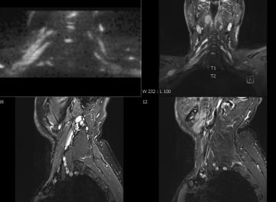
Current Concepts on MR Neurography
Synopsis
MR Neurography has stepped out from being an emerging technique into clinical retort routine. This lecture will review current concepts on MR neurography and will provide an impression on how it may be used on a day-to-day basis.
MR neurography is a term which refers to dedicated MR imaging of peripheral nerves. This includes the spine, the nerve roots, as well as the plexus and peripheral nerves. Almost all peripheral nerves can be visualized on high-resolution MR neurography images, if dedicated protocols are used, and if the nerves are not extremely small. Much research has been performed in the past years with a rapidly increasing knowledge about how to apply, optimize and perform MR neurography on the day to day basis for clinical routine. Frequent findings in MR neurography are of course directly related to the frequency of nerve abnormalities. Causes for nerve disorders include but are not exclusively limited to:
1. systemic diseases, such as ischemia, vasculitis, toxic, endocrine and metabolic disorders such as diabetes amyotrophy, hereditary motor-sensory neuropathies, amyloidosis, hyperlipidemia, acute and chronic demyelinating inflammatory neuropathies, etc.
2. local conditions, such as plexopathy, nerve injury, perineural compressive lesions, adhesive neuropathy in failed tunnel cases, infections and nerve sheath tumors, etc.
3. neuropathies related to functional anatomical changes, such as habitual leg crossing, repeated typing and repetitive exercise, which may cause traction mononeuritis or compressive neuropathy in functional compartment syndromes.
Some of the aforementioned disorders can perfectly be diagnosed on the clinical basis without imaging. However, in several of them, MR neurography will have a significant clinical impact on establishing the final diagnosis, as well as guiding therapy and follow-up. These has been shown by several studies. Nevertheless, the role of MRI in these conditions needs to be understand:
1. In systemic nerve diseases, MRI is not expected to make these diagnoses. However, MRI may be used to confirm the clinical suspicion by demonstrating abnormality of the innervating and regional muscle denervation changes or, exclude any structural cause for mononeuropathy in these cases.
2. In local conditions, MRI has a major role in these cases as it supplements information gained from the clinical exam and electrodiagnostic tests or provides information, which may not be possible to attain from other modalities.
3. In neuropathies related to functional anatomical changes, the role of MRI is limited. This group is mostly diagnosed on clinical basis and pressure catheter studies. However, rarely, MRI may be used in clinically confounding cases with imaging performed before and after the exercise/effort in question. Indirect signs such as significant prolongation of T2 signal intensity within the muscle compartment may aid in the diagnosis of compartment syndrome.
This talk will focus on current concepts for clinical application of MR neurography with a focus on standard sequences that are helpful in the clinical routine. New sequences such as such as simultaneous multislice imaging as well as nerve specific 3-D sequences will be discussed as well, since these types of sequences have become commercially available within the last year from different vendors. This talk will mention but not focus on diffusion tensor imaging and diffusion weighted imaging. These two techniques are still under development and much research is going on, especially in the terms of establishing standard values and normative parameters for clinical use.
