5650
1H NMR based metabolomics study of serum in Parkinson’s patients1Department of NMR and MRI Facility, All India Institute of Medical Sciences, New Delhi, India, 2Department of Neurology, All India Institute of Medical Sciences, New Delhi, India, 3Department of Biostatistics, All India Institute of Medical Sciences, New Delhi, India
Synopsis
Proton metabolic profile of serum samples in 20 patients with Parkinson’s disease (PD) and 10 healthy controls (HC) was studied using 700 MHz NMR spectrometer. Data were processed using MestReNova software (version: 10.0) and integral values were evaluated. PLS-DA multivariate analysis was performed to compare the metabolic differences between PD patients and HC using MetaboAnalyst (version: 3.0) software. We found elevated levels of glucose, fatty acid, glutamine, lactate, choline, creatine and acetate in PD patients in comparison with HC (on t-test, p<0.05), indicating disturbances in lipid metabolism, fatty acid oxidation and mitochondrial damage leading to dopaminergic deficiency in Parkinson's disease.
Purpose
To study the metabolic profile of serum sample by in vitro NMR spectroscopy and identify possible bio-markers for Parkinson's Disease.Introduction
Parkinson’s disease (PD) is the second most common neurodegenerative disorder, characterized by progressive loss of dopaminergic neurons in the substantia nigra pars compacta (SNpc)1. Proton nuclear magnetic resonance based metabolomic analysis is a powerful tool for the determination of biomarkers for neurodegenerative disease. We used 1H NMR technique with multivariate analysis to identify possible biomarkers in serum sample of PD patients which could help in diagnosis of the disease.
Methods
Blood samples were collected from PD patients (n = 20; 13M/7F; mean age: 57 ± 7.0 years) and 10 age and gender matched healthy controls (HC) (4M/6F; mean age: 49.0 ± 2.22 years) after 12 hours fasting, centrifuged at 2000 g for 10 min at 4o C and stored the supernatant sample at -80oC until the NMR experiment. We used 200 µl serum, 30 µl Sodium formate (0.5mM, used as reference) and D2O solvent (to making total volume 600µl) for NMR experiments. NMR spectra were acquired using 1D CPMG pulse sequence using a 700 MHz NMR spectrometer (Varian, M/s. Agilent Technologies, USA). Parameters were: scans 64; echo time 15 ms ; relaxation delay 70 s. Peaks were assigned using 1D, 2D spectra (zTocsy) and data were processed using MestReNova software (version10.0, Mestrelab Research, Spain). PLS-DA multivariate analysis were carried out using MetaboAnalyst (version:3.0), a web-based metabolomics data processing tool for comparison between PD patients and HC.Results
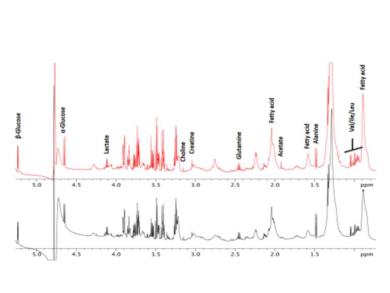
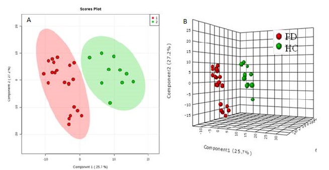
Fourteen metabolites (α, β-glucose, fatty acid (Lip09, Lip21), glutamine, lactate, choline, creatine, glutamine, acetate, alanine, leucine, isoleucine and valine) were assigned as shown in representative 1D NMR spectra (Figure 1) and integral values evaluated. Significant increase in glucose, fatty acid, glutamine, lactate, choline, creatine and acetate were observed in PD patients as compared to HC on t- test (non-parametric Wilcoxon rank-sum test, p<0.05). PLS-DA analysis plot depicts clear separation between PD and HC (Figure 2).Discussion
Significant differences were observed in metabolic profile of sera between PD patients and control group. Higher levels of lactate may be attributed to disturbances in energy and glucose metabolism2. Increased levels of glutamate may be ascribed to impaired mitochondrial function, due to increased vulnerability of affected neurons in PD as compared to HC3. Increased levels of Lip09, Lip21 may be due to enhanced lipid metabolism and fatty acid oxidation in PD. Presence of Lewy bodies in PD with characteristic inclusions of aggregated alpha-synuclein has been implicated in a range of interactions with phospholipid membranes and free fatty acids. Association of alpha-synuclein with oxidized lipid metabolites may lead to mitochondrial dysfunction in Parkinson's disease4. These results demonstrate that metabolic profiling of serum could be useful as a screening tool for early diagnosis and prognosis, and enhance our understanding of the mechanisms involved in the disease progression.Conclusion
Metabolomic analysis of serum sample identified potential biomarkers associated with lipid metabolism, energy and glucose metabolism which would help in understanding PD pathogenesis.Acknowledgements
No acknowledgement found.References
1. Lu, Z et al. 1H NMR-based metabolomics study on a goldfish model of Parkinson’s disease induced by 1-methyl-4-phenyl-1, 2, 3, 6-tetrahydropyridine (MPTP). Chemico-biological interactions. 2014; 223: 18-26.
2. Raquel D. et al. Oxidative stress and aminopeptidases in Parkinson’s disease patients with and without treatment. Neurodegenerative Diseases. 2011; 8: 109-116.
3. Beal, MF, Lang AE, and Ludolph AC. Neurodegenerative Diseases: Neurobiology, Pathogenesis and Therapeutics. Cambridge University Press, 2005; ISBN:9780511113741.
4. Ruipérez, V, Frédéric D and Bazbek D. Alpha-synuclein, lipids and Parkinson’s disease. Progress in lipid research. 2010;49: 420-428.
Figures