5649
Using broadband refocusing pulses for increased sensitivity for 2HG detection to determine glioma IDH mutation status1Memorial Sloan Kettering Cancer Center, New York, NY, United States, 2GE HealthCare, Berlin, Germany, 3GE Global Research, Munich, Germany
Synopsis
2-hydroxyglutarate in IDH1/2 mutated tumors is of clinical interest and can be measured in-vivo by magnetic resonance spectroscopy. Goal of this work was to compare results obtained using the standard reduced flip angle refocusing pulses with results using broadband refocusing pulses. Sensitivity improvement was observed in phantoms and demonstrated in vivo.
Purpose
The detection and quantification of 2-hydroxyglutarate (2HG) in IDH1/2 mutated tumors is of clinical interest because the elevated 2HG level has been linked to IDH1/2 gene mutation in glioma. 2HG can be measured in-vivo by MR spectroscopy [1,2] and might be used as a biomarker for both diagnosing IDH mutations in tumors as well as monitoring response to treatments targeting these mutations. The point-resolved spectroscopy (PRESS) technique is a commonly used technique on clinical systems and it has been shown that optimizations of subecho times (TE1/TE2 pair) [1] increase the sensitivity of 2HG detection. Goals of the present work is 1) To optimize the TE1/TE2 pair for the maximum sensitivity of 2HG using GE 3T MR scanners, 2) To apply broadband refocusing pulses [3] and comparing results with those using the standard reduced flip angle product pulses [4] at the optimal TE1/TE2 pair.
Methods
This is a prospective IRB approved HIPAA-compliant study with informed patient consent.
Experiments were performed both in a MRS phantom (13mmol 2HG, 20mmol Glycine, pH 6.8) and in-vivo in a 3.0T clinical scanner (MR750w, GE Healthcare).
S-BREBOP pulses (broadband and B1-robust refocusing with 2.8kHz bandwidth) [2] were implemented to replace the standard reduced flip angle (RFP) refocusing pulses (137°, 1.4kHz bandwidth; 167°, 1.1 kHz bandwidth) [3] of PROton Brain Exam using PRESS (PROBE-P). TE of 97ms (TE1 = 26ms, TE2 = 71ms) [1] was used. Center frequency was set to 2.7ppm (default).
All raw MRS data were processed with our in-house Matlab code to generate the coil-combined signal from eight channels using an unsuppressed water signal using 1-Hz gaussian filter. Further, this data was analyzed using LCModel fitting using exact Hamiltonian basis sets [2].
Results
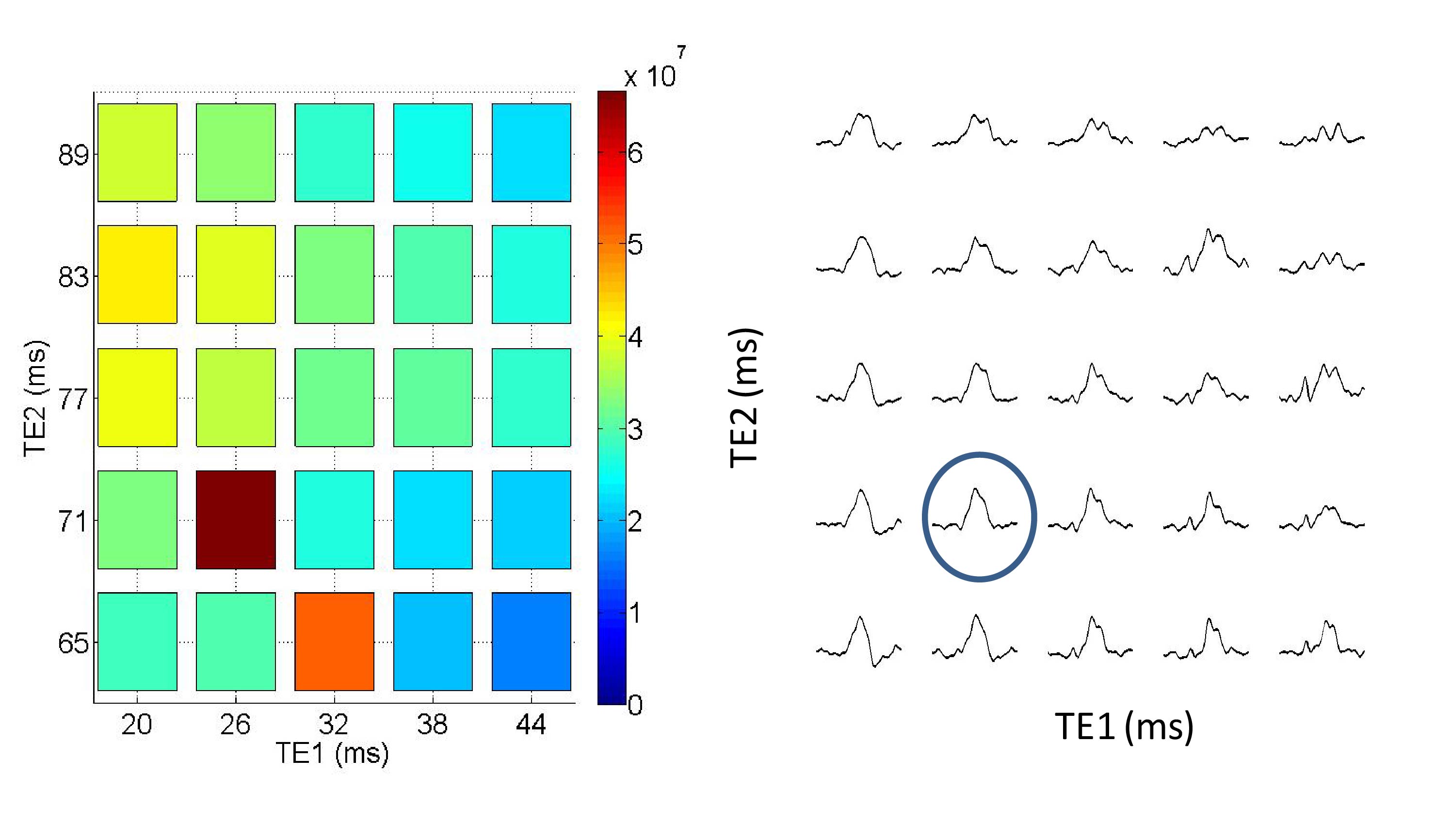
TE1/TE2 optimization using phantom: 2HG signal sensitivity is higher at the subecho times TE1=26ms and TE2=71ms (Figure 1-right panel). Integral was calculated from 2.2 to 2.3 ppm range to generate the color map (Figure 1-left panel).
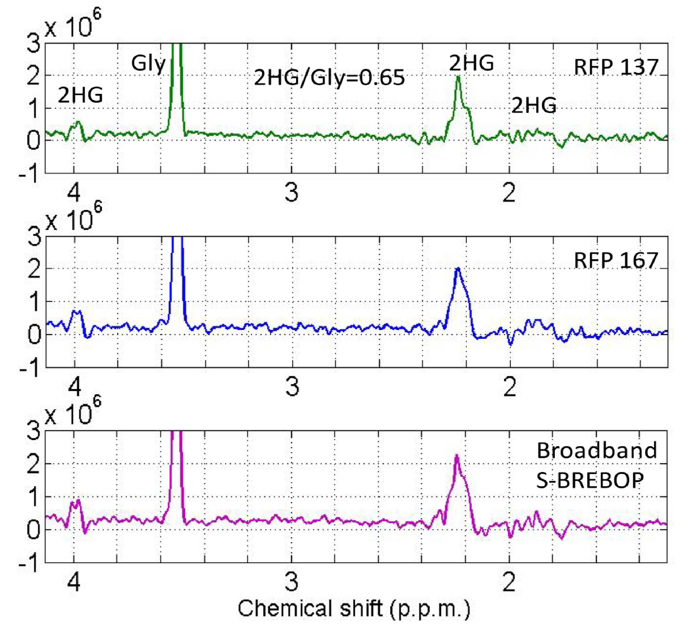
Comparison of broad band refocusing pulse with product RFP pulses: The largest 2HG signal at 2.25ppm is higher with the S-BREBOP pulses (Figure 2). The resonances between 1.8ppm and 2.0ppm and at 4.0ppm show large differences in terms of intensities and are lower in the acquisition using the standard reduced flip angle pulses (RFP 137 and RFP 167).
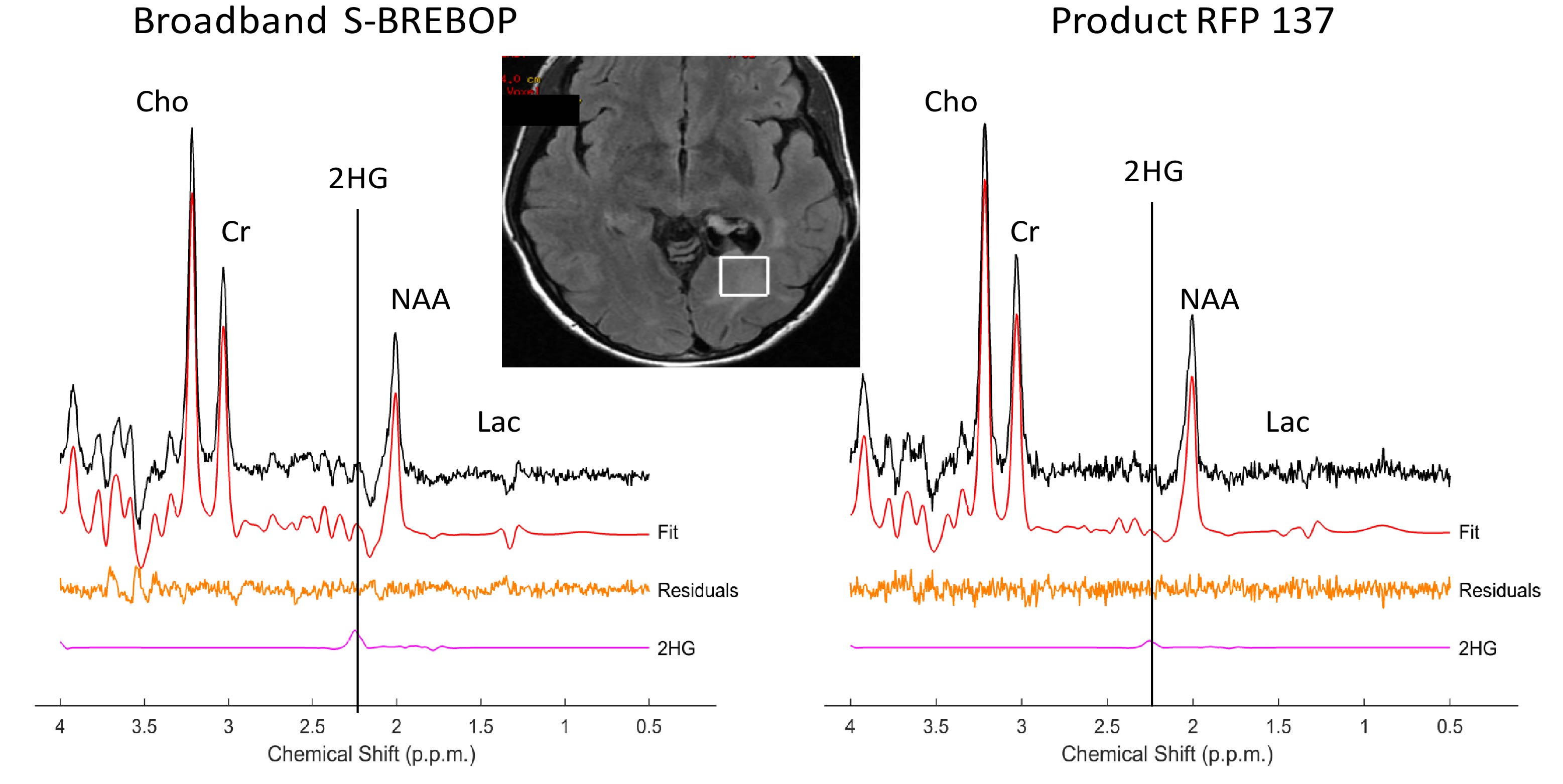
In vivo demonstration: The in vivo spectrum of 2HG acquired with the broadband S-BREBOP pulses show higher sensitivity compared with RFP 137 pulse routinely used for conventional 2HG MRS (Figure 3). The 2HG concentrations were calculated to be much higher (7.2 mM with CRLB=16%) using broad band pulse than the RFP 137 pulse.
Discussion
It has been shown that TE1=26ms and TE2=71ms can be used for optimal sensitivity of 2HG on GE scanners. It is also shown that the use of broadband refocusing pulses in a PRESS sequence increases the sensitivity for 2HG detection resulting in higher concentration values. The implementation of 2HG MRS on the different vendor systems especially the used RF pulses can have a significant influence on the measured concentrations.
Acknowledgements
Brain Tumor Center grant, B*Cured foundation grant, MSK society grant, and Dana Foundation grantReferences
Choi et al. Nat Med. 2012 Jan 26; 18(4): 624-9
de la Fuente et al. Neuro Oncol. 2016 Feb; 18(2):283-90.
Janich et al. J Magn Reson. 2012 Oct; 223: 129-37
Raidy et al. ISMRM 1995, 1020
Figures