5647
Investigation of metabolic changes during watching movie using fMRS at 3T system1Department of Electronic and Computer Engineering, National Taiwan University of Science and Technology, Taipei, Taiwan, 2Graduate Institute of Applied Physics, National Chengchi University, Taipei, Taiwan, 3Research Center for Mind, Brain and Learning, National Chengchi University, Taipei, Taiwan
Synopsis
Recent studies have reported metabolic change during visual and motor stimulation using MRS at 7T MRI system. We think there is potential to perform fMRS experiments at 3T system. Visual stimuli were given with block design consisting of 2 black-white movie clips and 3 rest sections. Our preliminary results showed that Glu concentrations in visual cortex increases by 1.39% during watching movie, while NAA and Cre have no significant change. The observed Glu change was comparable to previous study performed at 7T.
Introduction
Recent studies have reported metabolic change during visual and motor stimulation using MRS at 7T MRI system1,2. In these studies, glutamate (Glu) and lactate have been found to increase by 2% and 17% during stimuli using block design functional MRS (fMRS) experiments. These studies have been performed at 7T for the consideration of SNR where the Cramer Rao Lower Bound (CRLB) of Glu can be below 5%. Recently, we have found that MRS with TE of 40 ms can have CRLB for Glu at level of 7% at temporal resolution of 6 seconds at 3T system. Therefore, there is potential to perform fMRS experiments at 3T system. The aim of this study is to investigate the feasibility of fMRS experiments at 3T system using visual stimuli.Methods
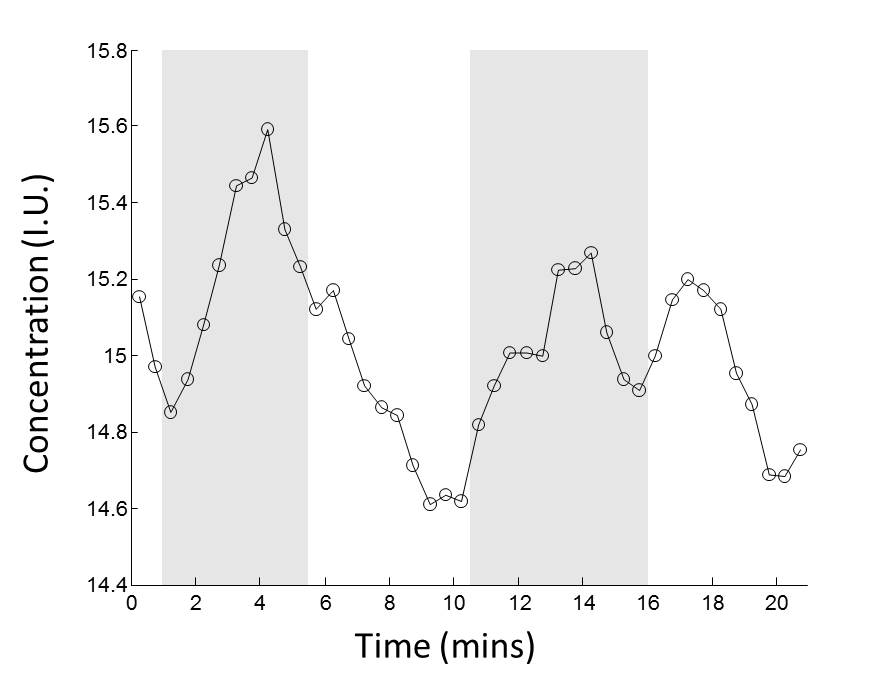
5 healthy subjects (age range: 22~27, 2 males and 3 females, normal or corrected to normal vision) were scanned using a 3T MR system (Skyra, SIEMENS Medical Solutions) with a 32-channels head coil. The 30x25x25 mm3 VOI was firstly defined on MNI template to cover most of visual cortex according to previous fMRI study3. For MRS scan, an automatic localization tool (ALLVOI)4 was used to locate the predefined VOI on T1 images of each subject to ensure consistency in spatial coverage of visual region among subjects. fMRS experiments were performed with parameters: TR/TE=1500/40 ms, bandwidth = 2000 Hz, vector size = 2048, 4 phase cycling steps. Visual stimuli were given by presenting black-white movie clips in 5 blocks: Rest (1:00), Movie1 (4:34), Rest (5:07), Movie2 (5:19), and Rest (5:00). Movie clip 1 was taken from 6’30” of the movie ‘‘The Circus’’ and Movie clip 2 was taken from 1:03’01” of the movie “City Light”. 210 measurements yield the total scan time of 21 minutes. A non-water suppression MRS was acquired using same parameters for water scaling. To increase spectral SNR, the acquired MRS spectra were averaged by a window of 5, which result in a total of 42 time points with temporal resolution of 30 seconds in the fMRS experiment. Acquired MRS data were quantified using LCModel. Metabolites concentrations of NAA, Cre and Glu were calculated using water scaling with partial volume and relaxation correction. The mean Glu, NAA and Cre concentrations during movie watching blocks section and during rest blocks were calculated for each subject. Paired student t-test was performed to evaluate the significance of the metabolic change at the presence of visual stimuli.Results
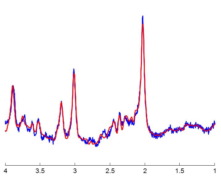
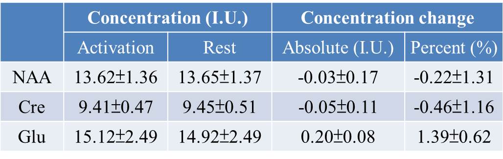
The representative spectrum from one time point (30 seconds) of fMRS experiment was shown in figure 1. The noise level is low enough to identify Glu peak on the spectrum by visual inspection and the CRLB of Glu for this spectrum is 7%, which stand for the overall CRLB levels 7.15%±0.56% for all subjects . The average CRLBs for NAA and Cre were 2.39%±0.31% and 2.28%±0.36%, respectively. Figure 2 showed the time course of Glu level averaged between subjects across fMRS scan. The change of Glu concentration along with paradigm can be observed. The mean Glu, NAA and Cre concentrations during movie watching blocks section and during rest blocks were listed in Table 1. Glu was significantly increased during watching movie by 1.39% (p<0.005). There was no significant change for NAA and Cre concentrations (p>0.1).Discussion and Conclusions
Our preliminary results showed that Glu concentrations in visual cortex increases by 1.39% during watching black-white movie, while NAA and Cre have no significant change. The observed Glu change was comparable to previous study performed at 7T1,2, which reported 2% increase in Glu level during visual and motor stimuli. The lower magnitude of change may be attributed to that we are using movie clip as visual stimulation instead of common checker board pattern, which is expect to excite intense visual stimulation. Our results also indicate that materials containing complex cognitive information can be used in fMRS experiments. The future goal of this study is to increase subject group to verify the feasibility of fMRS experiments at 3T system and based on the finding of this study we will use the same material to study the metabolic change in high order cognitive regions instead of primary sensory region.Acknowledgements
No acknowledgement found.References
1.Schaller, B., et al. (2014). "Are glutamate and lactate increases ubiquitous to physiological activation? A 1 H functional MR spectroscopy study during motor activation in human brain at 7Tesla." Neuroimage 93: 138-145.
2.Lin, Y., et al. (2012). "Investigating the metabolic changes due to visual stimulation using functional proton magnetic resonance spectroscopy at 7 T." Journal of Cerebral Blood Flow & Metabolism 32(8): 1484-1495.
3.Jaaskelainen, I. P., et al. (2016). "Brain hemodynamic activity during viewing and re-viewing of comedy movies explained by experienced humor." Sci Rep 6: 27741.
4.Peng Po-Yu, et al. "Toolbox for automatic localization of volume of interest in MRS (ALLVOI) ". ISMRM 2016; Abstract ID: 3981.
Figures