5646
Altered intrinsic neuronal activity correlates with GABA levels in the auditory region of patients with presbycusis1Shandong Medical Imaging Research Institute, Shandong University, JINAN, People's Republic of China, 2Philips Healthcare, Shanghai, People's Republic of China
Synopsis
In this study, we used J-difference edited MRS and resting-state fMRI to investigate correlation between intrinsic neuronal activity and GABA levels in the auditory cortex of patients with presbycusis. Our results indicated that abnormalities in GABAergic neurotransmission may underlie resting-state functional deficits in presbycusis.
Purpose
Presbycusis is the most common sensory deficit in the ageing population. Patients with presbycusis suffer not only from hearing loss, but also from poor speech discrimination, which reflects functional changes of the auditory cortex. Decreased levels of gamma-aminobutyric acid (GABA), the main inhibitory neurotransmitter in the central auditory system, have been found in the auditory cortex in patients with presbycusis using magnetic resonance spectroscopy (MRS).1 Previous studies have demonstrated that GABA is involved in modulating intrinsic neural activity.2 However, to date there has been a paucity of studies exploring correlation between intrinsic neuronal activity and GABA levels in the auditory cortex of patients with presbycusis. In this study, therefore, J-difference edited MRS and resting-state functional magnetic resonance imaging will be used to investigate the neurochemical mechanism of presbycusis.Methods
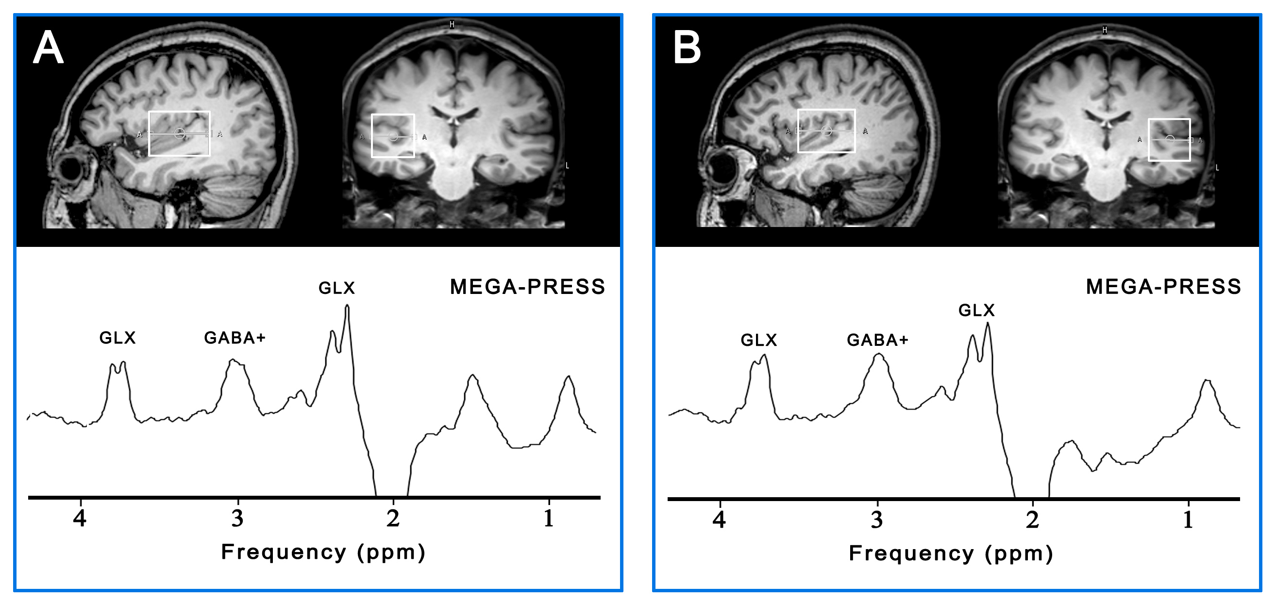
Fifteen patients (right-handed) with presbycusis (5 males/10 females, mean age 63.2 ± 2.6 years) and thirteen age- and gender-matched healthy controls (5 males/8 females, mean age 62.2 ± 1.9 years) were recruited in this study. All experiments were performed on a 3T scanner (Philips ‘Achieva’ TX, Best, The Netherlands) equipped with a 8-channel head coil. The hearing abilities of all subjects were evaluated using pure tone audiometry and tympanometry. The MEGA-PRESS sequence was used to measure GABA levels in 4 x 3 x 3 cm3 volumes centered on the left and right Heschl’s gyri (HG), as shown in Fig. 1. The MEGA-PRESS data were analyzed using 'Gannet' (GABA-MRS Analysis Tool) in Matlab with Gaussian curve fitting to the GABA peaks.3 The rs-fMRI data was analyzed with SPM and REST in Matlab. The fALFF within the MRS voxel was obtained using the ratio of power spectrum in a given frequency band (0.01–0.08 Hz) to the total power in the entire detectable frequency range (0.009–0.25 Hz).Results
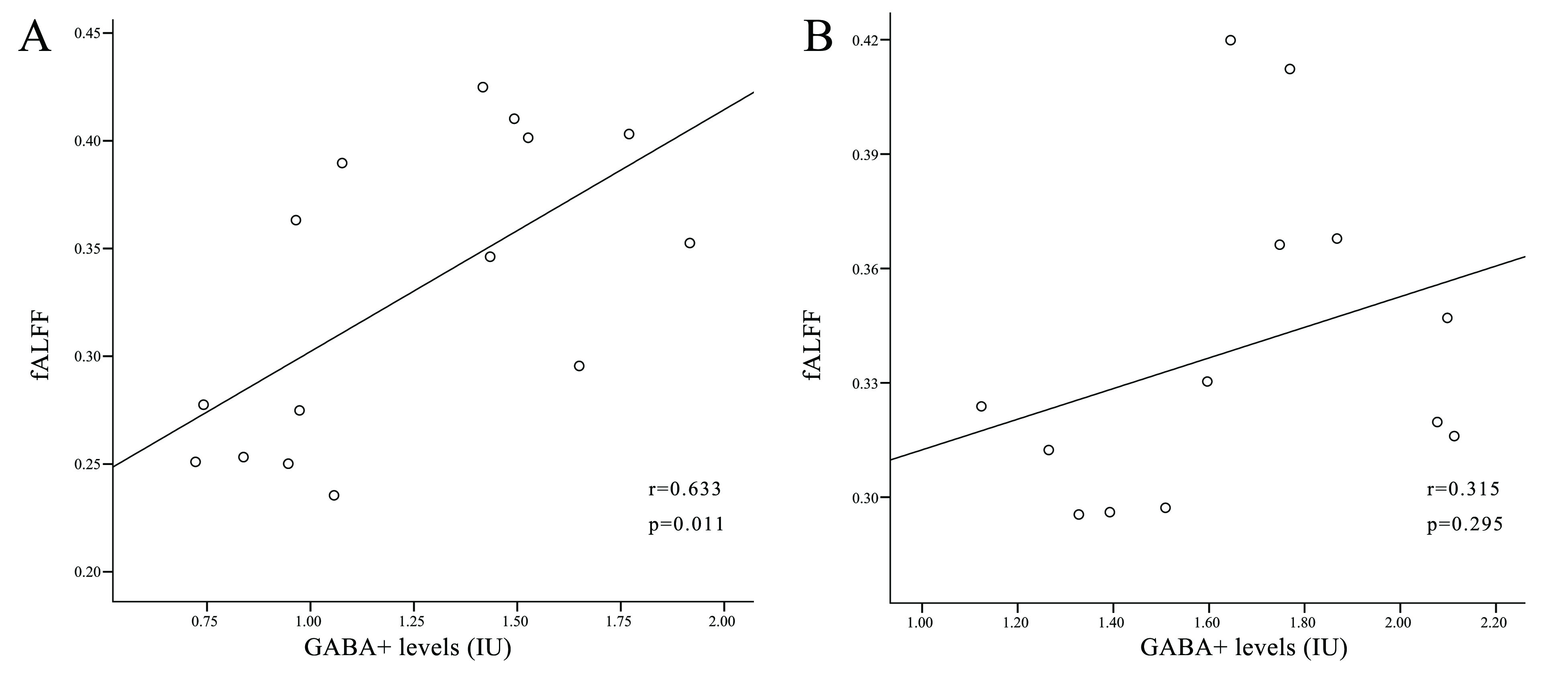
For healthy controls, there were no correlations between GABA levels and fALFF in the left (r = 0.315, p = 0.295) and right MRS voxel (r = -0.13, p = 0.672); For patients with presbycusis, significant positive correlations were observed between GABA levels and fALFF in the left MRS voxel (r = 0.633, p = 0.011) (Fig. 2); there was no correlation between GABA levels and fALFF in the right MRS voxel (r = 0.095, p = 0.737).Discussion
In our study, significant positive correlations between GABA levels and fALFF were observed in the left auditory region of patients with presbycusis, while these correlations were not found in healthy controls. GABA plays an important role in the modulation of small scale synchrony of microcircuits, which correlates with the level of whole-brain distributed networks.2 Previous studies have demonstrated there is a dysfunctional GABAergic neurotransmission in the central auditory system in presbycusis.1, 4 Our results indicated that abnormalities in GABAergic neurotransmission may underlie resting-state functional deficits in presbycusis.Conclusion
It is believed that our findings could be important for exploring associations of regional GABA levels with intrinsic neural activity. The multimodal resting-state neuroimaging technique served as a useful tool for functional-biochemical investigation of presbycusis.Acknowledgements
This project was supported by the National Natural Science Foundation of China (grant nos. 81601479) and Shandong Provincial Natural Science Foundation of China (grant nos. BS2015YY003) and Shandong Provincial Medical and Healthy Technology Development Program of China (grant nos. 2015WS0176).References
1. Gao F, Wang G, Ma W, et al. Decreased auditory GABA+ concentrations in presbycusis demonstrated by edited magnetic resonance spectroscopy. NeuroImage 2015;106:311-316.
2. Duncan NW, Wiebking C, Northoff G. Associations of regional GABA and glutamate with intrinsic and extrinsic neural activity in humans-a review of multimodal imaging studies. Neurosci Biobehav Rev 2014;47:36-52.
3. Edden RA, Puts NA, Harris AD, et al. Gannet: A batch-processing tool for the quantitative analysis of gamma-aminobutyric acid-edited MR spectroscopy spectra. J Magn Reson Imaging 2014;40:1445-1452.
4. Syka J. The Fischer 344 rat as a model of presbycusis. Hear Res 2010;264:70-78
Figures