5640
Decreased tNAA concentration in female college basketball players with mild depression/anxiety symptoms1Psychiatry, University of Utah, Salt Lake City, UT, United States
Synopsis
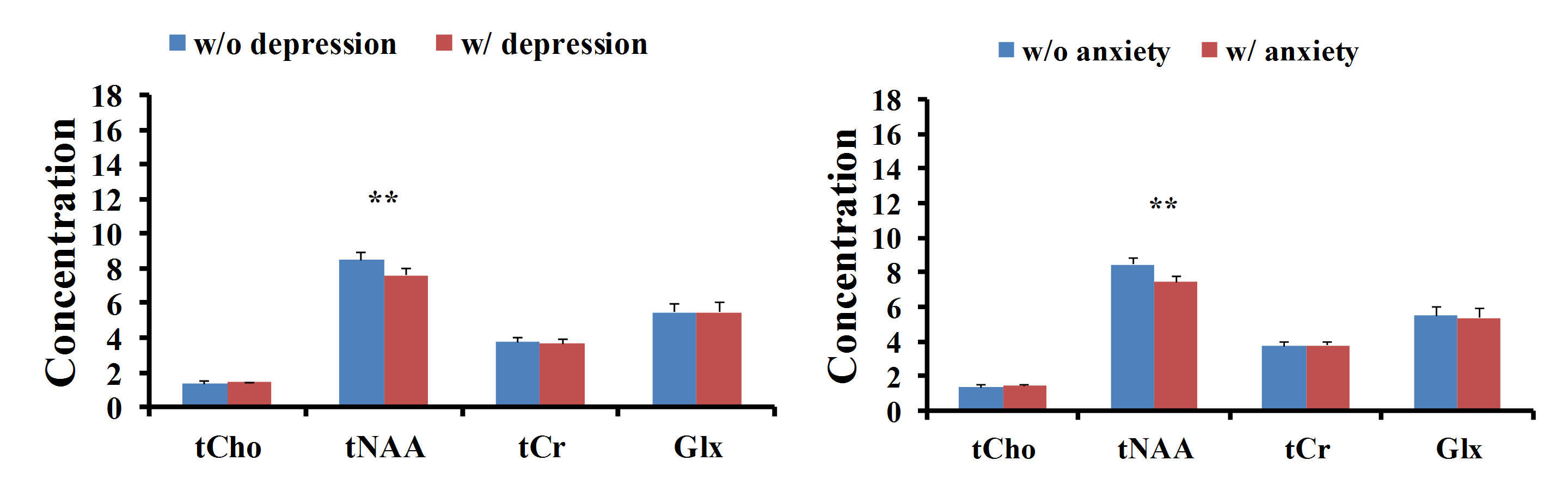
The aim of the present study was to examine possible gender differences in 1H brain metabolite concentrations in male and female college basketball players. Decreased total N-acetylaspartate/N- acetylaspartylglutamate (tNAA) levels within white matter tissue were observed in female basketball players with symptoms of depression or anxiety (p = 0.0256 /p = 0.0112).
INTRODUCTION:
Depression and anxiety are two common disorders that negatively effect social functioning. A commonly held belief about athletes is that physical activity and athletic participation may lower risk for depression. However, Wolanin et al.1 recently reported that the prevalence rate of depression among college athletes is 23.7% with female athletes of 28.5%. These rates are much higher than the 12-month depression prevalence rates of 6.7% among US adults. Further, the estimated lifetime prevalence of major depression is greater in women (11.7%) than in men (5.6%). Anxiety also often co-occurs with depression. In previous studies2, magnetic resonance spectroscopy has been used to produce the neurochemical consequences of sport-related concussion among college athletes. However, brain imaging studies of depression and anxiety among college athletes are understudied.METHOD:
16 male (age =20.1±1.0 years; age range = 18-22 years) and 14 female (age = 20.3±1.3 years; age range = 18-22 year) divisional varsity college basketball players were enrolled. The study protocol was approved by the University Institutional Review Board, and written informed consent was obtained from all subjects. All scans were acquired on a 3 Tesla (T) clinical Verio MRI system. 1H spectra were acquired using a 2D CSI pulse sequence with TR/TE 1590/40 ms, receiver bandwidth 1 kHz, Field of View 22x22x1.5 cm3, Volume of Interest (VOI) 8x7x1.5 cm3, matrix scan size 24x24, the nominal voxel size 6.875x6.875x15 mm3 after zero filling in k-space to 32x32 samples, and vector size 1024. To facilitate voxel placement, high resolution T1 weighted images were acquired using a MPRAGE pulse sequence. The VOI covers brain regions encompassing frontal lobe and anterior cingulate cortex and situates immediately above the corpus callosum (Fig. 1). To evaluate the influence of brain tissue components within the VOI, MPRAGE images were segmented into gray matter (GM), white matter (WM) and cerebrospinal fluid (CSF) using FSL. After zero-filling to 32x32 points in k-space, applying a Hamming filter with a 50% window width, and 2D spatial Fourier transformation, the time domain 2D CSI data were analyzed using LCModel. Poor data exclusion criteria consisted of the Cramer-Rao lower bounds of the fit to the peak of interest (> 20%). Estimates of metabolite concentrations in either GM or WM were computed by linear regression of the CSF-corrected metabolite concentration in each, against the normalized GM fraction of the voxel, and extrapolating to a GM fraction of one (pure GM), or zero (pure WM). The CSF-corrected metabolite concentration data were then divided into two groups by gender. To study metabolite concentration difference in subjects with/without symptoms of depression/anxiety, two subgroups were defined by subjects with HAM-D/A scores (> 0 or = 0). Finally, the gender as a co-factor appears in the final subgroup data analysis in order to investigate gender difference in the subgroup data. Statistical analyses were performed using R statistics software. Unpaired student’s t-test was applied to group data. P values were adjusted by post-hoc Bonferroni correction to reduce family-wise error. A Bonferroni corrected p value of < 0.05 was considered to be statistically significant difference.RESULT & DISCUSSION:
No group differences of metabolite concentration by gender were observed in pure gray matter and pure white matter, respectively. However, the subgroup data analysis shows that white matter tNAA concentration in subjects with symptoms of depression/anxiety (HAM-D/A score > 0) is significantly lower than that in subjects without symptoms of depression/anxiety (HAM-D/A scores = 0). After the gender is introduced as a co-factor in the subgroup data analysis, a significant relationship between tNAA level and Hamilton score is noted in female basketball players. This significant metabolite concentration difference appears in white matter tissue instead of gray matter tissue (Fig. 2) in female basketball players. As we know, the brain tNAA is present predominantly in neuronal cell bodies, where one of its roles is acting as a neuronal density marker. Altered neuronal density may reflect disturbed mitochondrial function. However, in the present study, the reduction of white matter tNAA level in female college basketball player with symptoms of depression or anxiety is noted when compared with those without symptoms of depression/anxiety. Our finding is consistent with the previous report that female athletes exhibited increased risk for depressive symptoms. In addition, Chamard et al.3 have found that female ice hockey players without concussion demonstrate decreased tNAA level over the course of season. The exact mechanism that underlies this finding is not clear. It may result from player depression or anxiety in highly competitive sports such as basketball players or ice hockey players.Acknowledgements
No acknowledgement found.References
[1] Wolanin, A., et al., Prevalence of clinically elevated depressive symptoms in college athletes and differences by gender and sport. Br J Sports Med, 2016. 50(3): p. 167-71.
[2] Gardner, A., G.L. Iverson, and P. Stanwell, A systematic review of proton magnetic resonance spectroscopy findings in sport-related concussion. J Neurotrauma, 2014. 31(1): p. 1-18.
[3] Chamard, E., et al., A prospective study of physician-observed concussion during a varsity university hockey season: metabolic changes in ice hockey players. Part 4 of 4. Neurosurg Focus, 2012. 33(6): p. E4: 1-7.
Figures