5634
Abnormal developmental trajectories of brain metabolites contributed to abnormal muscle tone development in infants with prenatal methamphetamine and tobacco-exposure1University of Hawaii at Manoa, Honolulu, HI, United States, 2Pediatrics, Sorlandet Hospital, Arendal, Norway
Synopsis
In prior studies, children with prenatal methamphetamine-(PME) or tobacco-exposure (PTE) showed elevated brain metabolites levels. The current study evaluated infants with PME and PTE during the first 5 months of life and found abnormal developmental trajectories of metabolites in the frontal white matter, with abnormally lower levels of total creatine [tCr], N-acetylaspartate [NAA], and glumate+glutamine [Glx] at baseline, and steeper developmental trajectories that resulted in normal or elevated levels after 2-months old. Furthermore, the trajectories of basal ganglia-[NAA] and corticospinal tract-[tCr] further contributed to the slower muscle tone development in PME infants, especially the males.
INTRODUCTION
Methamphetamine (Meth) is the second-most commonly abused category of illicit drugs worldwide.1 Although 70-90% of Meth-users smoke tobacco cigarettes concurrently,2 the impact of comorbid Meth and tobacco-use during pregnancy on fetal brain development is rarely studied. Therefore, we evaluated whether the developmental trajectories of brain metabolites are altered in neonates with prenatal Meth+tobacco-exposure (PME) or prenatal tobacco-exposure (PTE) during the first 5 months of life using 1H-MR spectroscopy (1H-MRS).METHODS
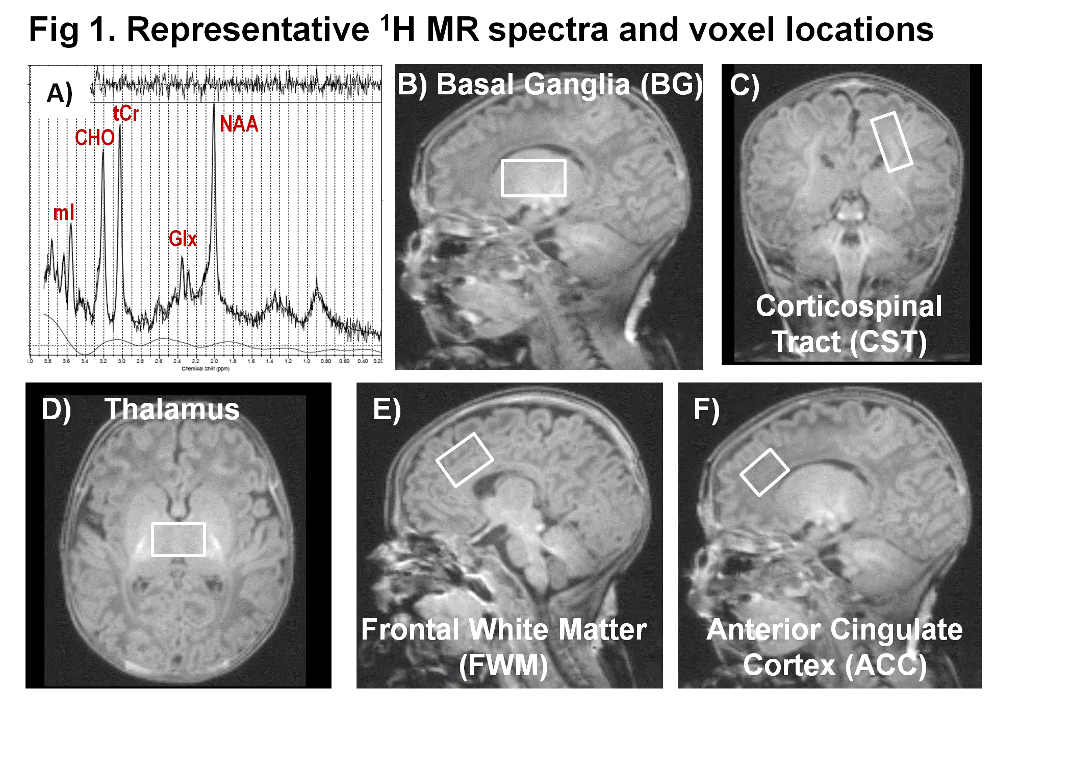
170 healthy neonates [85 unexposed (52.9% female), 42 PME (57.1% female), 43 PTE (34.9% female)] fulfilling study criteria were scanned 1-5 times over the first 5 months at 3 Tesla (Siemens TIM Trio). 1H-MRS was performed using PRESS (TR/TE=3000/30ms) in 5 brain regions (Fig. 1), measuring the T2 decay of water at 10 TE-values to correct for CSF partial volumes. Data were processed using LCModel3 to determine concentrations of N-acetyl aspartate (NAA), choline (Cho), total creatine (tCr), glutamate +glutamine (Glx), and myo-inositol (mI). The infants were also evaluated serially with Amiel-Tison Neurological Assessment at Term (ATNAT). Mixed model repeated-measures ANCOVAs were performed to assess group differences in metabolite development and ATNAT scores. Postmenstrual age (PMA), PMA2, sex, and Index of Social Position (ISP) were included as covariates. P<0.05 were considered significant for group effects and group interactions.RESULTS
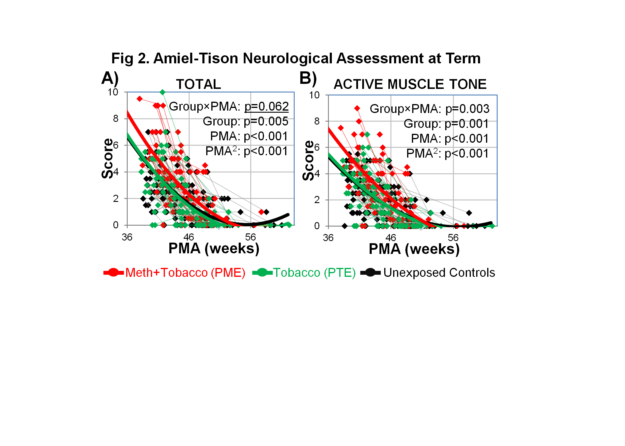
Participant characteristics: The mothers had similar ages at delivery (PME: 28.5±1.0 years; PTE: 26.3±0.8 years; Unexposed: 28.3±0.7 years), but PME mothers had greater pregnancy weight gain (20.5±1.6kg) than PTE (14.2±1.6kg) and Unexposed (14.4±0.7) mothers, p<0.0007. Additionally, compared to Unexposed mothers, PME and PTE mothers had lower index of social position (ISP: 63.7±1.0 and 52.7±2.1 vs. 44.2±1.6; p<0.001). During pregnancy, PME mothers cumulatively used 78±16g Meth and smoked more tobacco cigarettes (2,250±360) than PTE mothers (1,190±200). At birth, the three neonate groups were similar in gestational ages (PME: 39.1±0.2weeks; PTE: 39.2±0.2weeks;Unexposed: 39.3±0.2weeks), but the PME group had lower BMI (12.3±0.2) compared to PTE (12.9±0.2) and Unexposed (13.1±0.1); p=0.01. At baseline imaging, the neonates were similar PMA (PME:42.9±0.7weeks; PTE:42.1±0.5weeks; Unexposed:41.5±0.3weeks), p=0.09. The baseline ATNAT showed poorer total scores and weaker muscle tone in the PME neonates (4.0±0.4) compared to PTE (2.9±0.3) and Unexposed neonates (3.0±0.1), p=0.02, but these group differences were no longer significant by 3-months of age (Fig. 2).
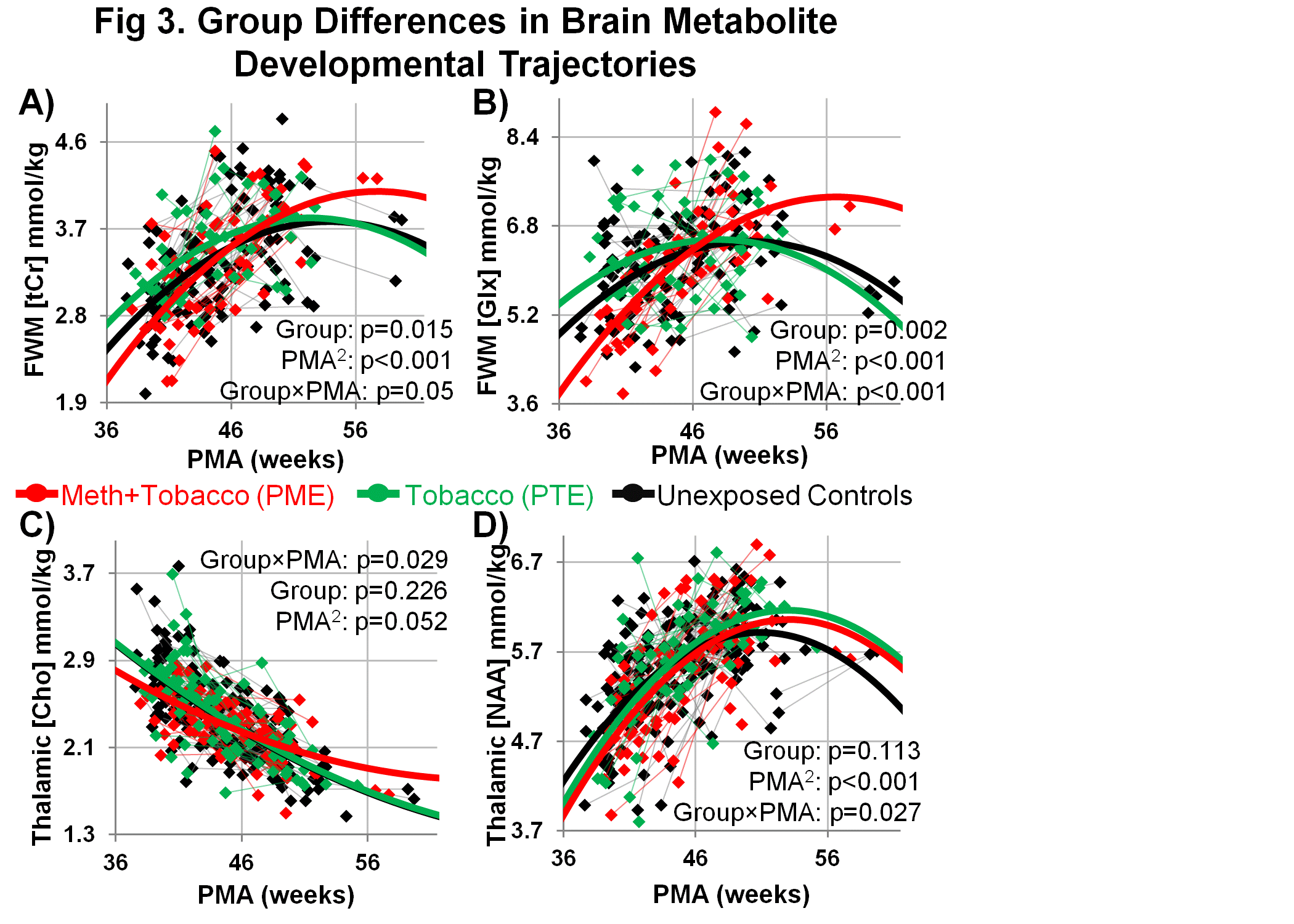
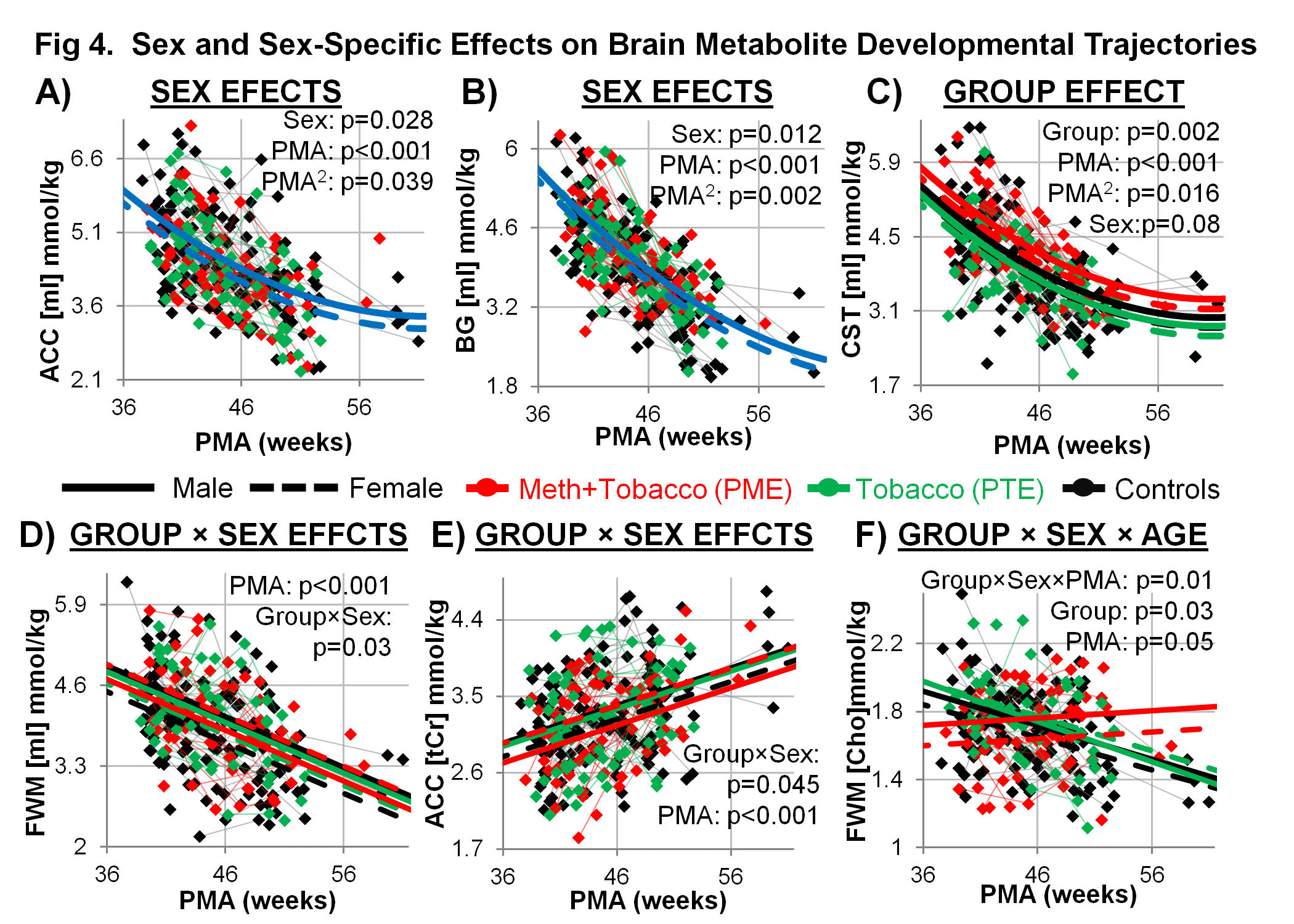
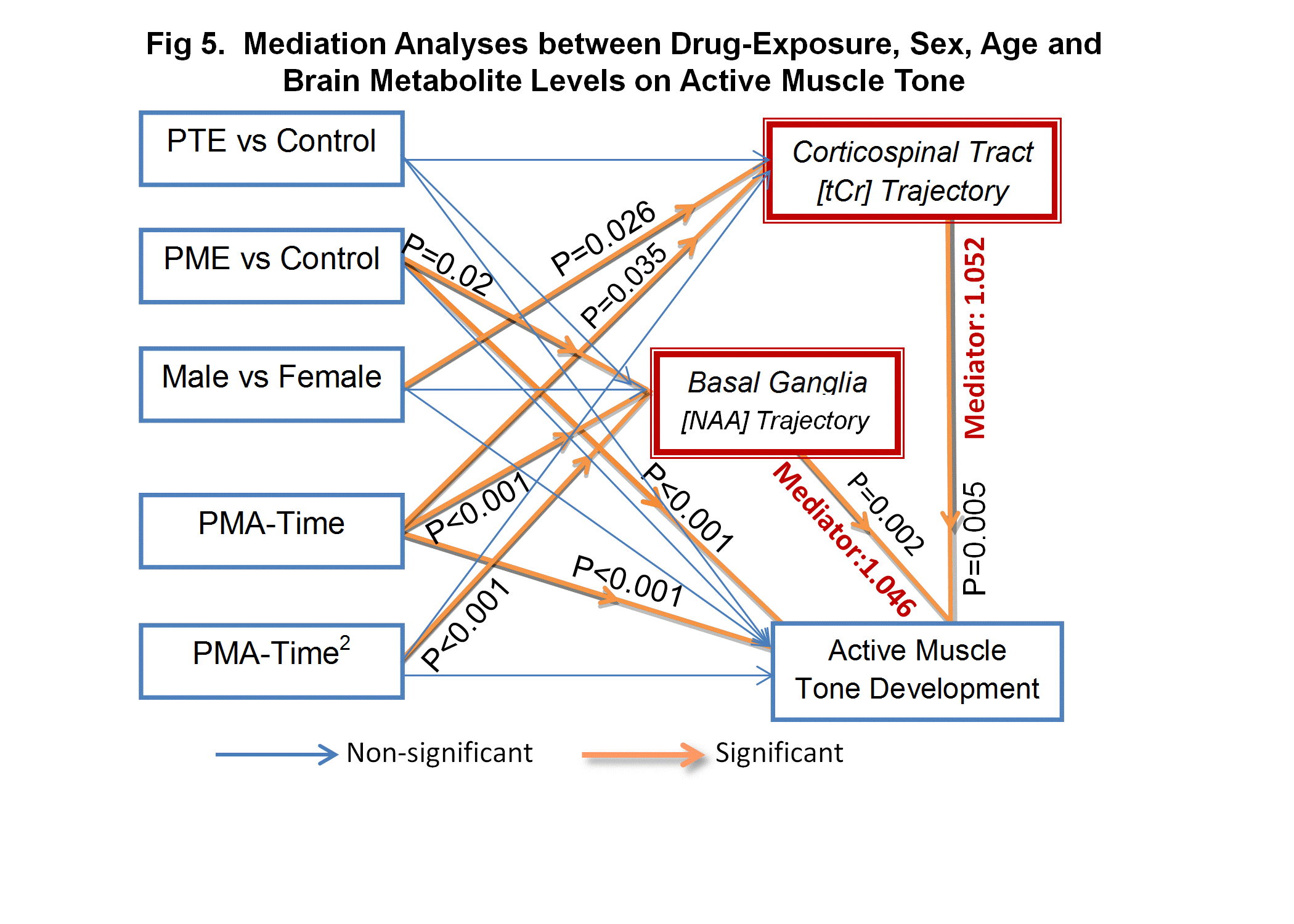
1H-MRS: Most brain metabolites showed age-dependent changes (p<0.05–0.001); therefore, PMA was included in final models. Fig. 3 shows that compared to PTE and Unexposed neonates, PME neonates had lower initial levels of FWM-[tCr], FWM-[Glx], thalamic-[Cho], and thalamic-[NAA]. However, these metabolite values normalized or increased to above normal after 48 weeks PMA. In Fig. 4, [mI] was lower in girls than boys in both BG (Sex: p=0.01) and ACC (Sex: p=0.03). CST-[mI] was lowest in PTE and highest in PME infants (Group: p=0.002) and tended to be lower in girls than boys (Sex: p=0.08). Additionally, group effects differed between sexes for FWM-[mI] (Group×Sex:p=0.03) and ACC-[tCr] (Group×Sex: p=0.045). Furthermore, PME infants showed altered development of FWM-Cho] in a sex-specific manner, with lower levels initially and lack of normal age-dependent decreases (Group×Sex×PMA: p=0.014; Group: p=0.034). Lastly, BG-[NA] further mediated the effects of PMA and PME on the abnormal muscle tone development by a multiplicative factor 1.046 (95%CI:1.004,1.110), while CST-[tCr] mediated the gender effect (being male) by a multiplicative factor 1.052 (95%CI:1.003,1.130), Fig. 5.
DISCUSSION:
During the first few months of life, the brain grows rapidly4,5 and our infants showed rapid age-dependent changes in brain metabolite concentrations. Although PME infants showed abnormally low levels of [tCr], [NAA], and [Glx] in FWM at baseline, their steeper developmental trajectories resulted in normal or elevated levels after 2-months old. The elevated brain metabolites at older age and the sex-specific effects are consistent with those found in prior 1H-MRS studies of PME children,6 including those at 3-4 years of age.7 These findings also complement altered DTI measures in PME infants,5 and are consistent with preclinical studies of prenatal Meth or Nicotine-exposure that led to aberrant axonal development with increased spine densities and altered myelin gene-expression.8,9 Collectively, our findings suggest that PME, both Meth and tobacco, was associated with altered neonatal neuronal and glial development, which might have resulted from epigenetic influences such as those found in rodents with prenatal Meth-exposure10 or prenatal nicotine-exposure.11 Finally, the abnormal BG-[NA] development contributed to the delayed muscle tone development in PME infants, while the gender effect mediated through CST-[tCr] slightly delayed the muscle tone development for boys. Longitudinal follow-up evaluations of our cohort are needed to better understand the impact of prenatal drug exposure on neurological development.Acknowledgements
Grant support from the NIH (U54-NS56883, K24-DA16170, K02-DA16991, G12-MD003061) and ONDCP (DABK39-03-C-0060); infrastructure support from the Queen’s Medical Center. We also thank our research participants, as well as the many technical and clinical staff at the Neuroscience and MR Research Program at the John A. Burns School of Medicine for their assistance with data collection and processing.References
1) United Nations Office on Drugs and Crime 2014, sales No. E.14.XI.7. 2) Weinberger AH & Sofuoglu M. Amer J Drug Alcohol Abuse 2009;35(1):12-17. 3) Provencher S. Magn Reson Med 1993;30:672. 4) Holland D et al. JAMA Neurology 2014; 71(10):1266-1274. 5) Chang L et al. JAMA Psychiatry 2016 Epub. 6) Smith L et al. Neurology 2001;67:255-260; 7) Chang L et al. NeuroImage 2009; 48(2):391-397. 8) Mychasiuk et al. Brain Res 2013;1499:53-60. 9) Cao et al. Transl Psychiatry 2013;3:e247. 10) Itzhak et al. Mol Psychiatry 2015;20(2)232-239. 11) Jung et al. Nat Neurosci 2016;19(7):905-914.Figures