5632
Validation and Initial Results from Dynamic 23Na fMRI1Aix-Marseille Université, Marseille, France, 2Heidelberg University
Synopsis
In this abstract we develop and validate an MRI acquisition/reconstruction method to derive the temporal dynamics of 23Na within a 20 min scan.
Introduction
Dynamic changes in intracellular Na+ act as signaling elements in the brain [1]. Prominent decreases in extracellular Na+ have been demonstrated in situ in response to glutamate or excitatory activity using sodium-selective microelectrodes [2,3]. Non-invasive monitoring techniques for Na+ changes are not presently suitable for observing changes on the time-scale of tens of seconds. Typical 23Na MRI acquisitions require 10-20 min to attain adequate SNR and spatial resolution.
In this work we develop and validate an MRI acquisition/reconstruction method to derive the temporal dynamics of 23Na within a 20 min scan. It is based on a short TE 3D-radial sequence and weighted least squares reconstruction using temporally dependent weights. The general approach is similar to the family of techniques such as sliding window, TRICKS [4] and VIPR [5], that share high spatial frequency k-space data.
Validation of the temporal dynamics was performed in phantoms using flip angle changes to mimic temporal variations during the scan. Feasibility and initial results in human subjects were demonstrated using a finger tapping task.
Experimental
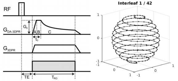
All experiments were performed on a Siemens 7T Magnetom using a dual tuned QED H/Na birdcage coil. A spoiled gradient echo, density adapted 3D projection reconstruction (DA-3DPR) 23Na MRI sequence was used with TR/TE = 120/0.2, BW 280 Hz/pixel, density adapted (t0 = 500 us), 10080 spokes [6]. Successive spoke angles were incremented by the golden angle and interleaved by a factor of 42 to give a smooth azimuthal variation through k-space and full polar coverage every 28.8 sec (see Figure 1) [7].
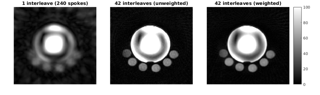
Reconstruction of one individual interleave (240 spokes) produced unusably blurred images (see Figure 2). Therefore a nonuniform Fourier transform reconstruction was implemented that uses weighted least squares to effectively share the high spatial frequencies from all interleaves but use the low spatial frequencies from a single interleave. To validate the approach, phantom experiments were performed using different flip angles on alternate interleaves (30 deg and 20 deg) in order to mimic signal activation. To provide a reference standard for the true signal, acquisitions were acquired at 30 deg and 20 deg separately.
For the in vivo study, one healthy volunteer (male, age 25yo) was recruited and in vivo measurements were taken during a finger tapping task 23Na fMRI. The time series of Na images (composed of 20 consecutive rest-activation periods: 30 sec active / 30 sec rest) were post-processed using statistical parametric mapping software (SPM12) with conventional procedures using realignment, spatial normalization smoothing (FWHM = 8 mm). The hand motion paradigm was entered in a general linear model (GLM) without haemodynamic response function (HRF) modeling.
Results
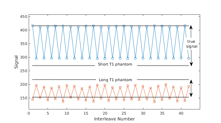
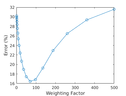
Phantom validation experiments produced time series that tracked the flip angle changes (see Figure 3). Note that the short T1 and long T1 phantoms signals move in opposite directions in response to the change in flip angle, so this is not a simple image scaling effect. A weighting factor of 100 was found to give the best compromise of contrast, resolution and artefact (Figure 4). In relation to the true signal measured from separate acquisitions, approximately 80% of the available signal variation was captured by the technique.
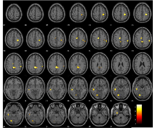
In vivo results during right hand movement produced significant patterns of Na decrease (t-test, p<0.005, k=10 uncorrected) during activation corresponding to the right hand motor system (see Figure 5).
Discussion
This study has used a weighted least squares approach to data-sharing in the context of dynamic 23Na MRI. The scheme produced temporal resolutions of 30 sec in 23Na images over the course of a 20 min scan. A recent study based on compressed sensing reported 64 sec temporal resolution for 23Na imaging [8]. The weighted least squares and compressed sensing approaches are complementary and may potentially be used in combination.
Validation using flip angle changes during the scan to mimic temporal variation made it possible assess the sensitivity of the technique. While there is some unavoidable blending of the contrast when sharing data, it was found that 80% of the available signal change could be captured with suitable parameter choices.
In application to the dynamics of 23Na in vivo, this study has shown a first proof of concept for the observation of extracellular sodium dynamics (a decrease in concentration) during cortical activation in an healthy control.
Acknowledgements
No acknowledgement found.References
1. Christine R. Rose and Alexei Verkhratsky. Principles of Sodium Homeostasis and Sodium Signalling in Astroglia. GLIA 2016;64:1611–1627
2. I. Dietzel, U. Heinemann, G. Hofmeier, and H.D. Lux. Stimulus-induced Changes in Extracellular Na + and CI- Concentration in Relation to Changes in the Size of the Extracellular Space* Exp Brain Res (1982) 46:73-84
3. Haack N, Durry S, Kafitz KW, Chesler M, Rose C. Double-barreled and Concentric Microelectrodes for Measurement of Extracellular Ion Signals in Brain Tissue. J Vis Exp. 2015 Sep 5;(103). doi: 10.3791/53058
4. J. Du, T.J. Carroll, H.J. Wagner, K. Vigen, S.B. Fain, W.F. Block, F.R. Korosec, T.M. Grist, and C.A. Mistretta. Time-Resolved, Undersampled Projection ReconstructionImaging for High-Resolution CE-MRA of the Distal Runoff Vessels. Magnetic Resonance in Medicine 48:516 –522 (2002)
5. C.A. Mistretta. Undersampled radial MR acquisition and highly constrained back projection (HYPR) reconstruction: Potential medical imaging applications in the post-Nyquist era. J Magn Reson Imaging Volume 29, Issue 3 March 2009 Pages 501–516
6. Armin M. Nagel, Frederik B. Laun, Marc-Andre ´ Weber, Christian Matthies,Wolfhard Semmler, and Lothar R. Schad. Sodium MRI Using a Density-Adapted 3D Radial Acquisition Technique. Magnetic Resonance in Medicine 62:1565–1573 (2009)
7. Bydder M. Mark Bydder, Wafaa Zaaraoui, and Jean-Philippe Ranjeva. Comparison of methods for choosing radial spoke directions in 3D UTE. ISMRM 2016:4312
8. Nicolas G.R. Behl, Armin M. Nagel, Erik N.K. Cressman, Reiner Umathum, David Fuentes, R. Jason Stafford, Peter Bachert, Mark E. Ladd, and Florian Maier. Time-resolved 23-Na Imaging for Monitoring of Thermochemical Ablation Injections. ISMRM 2016:199
Figures