5627
Simultaneous multi-parametric and quantitative estimation of 23Na physical properties at 7 Tesla using QuICS1Neurospin, CEA, Pais-Saclay University, Saclay, France, 2CRMBM, UMR 7339, Aix-Marseille University, Marseille, France, 3Institut Cerveau Moelle - ICM, CENIR, UPMC-Inserm U1127, CNRS 7225, Paris, France, 4Siemens Healthineers, Saint-Denis, France, 5IRFU, CEA, Paris-Saclay University, Saclay, France
Synopsis
Quantifying physical properties of sodium could be of benefit to assess more specifically changes in cellular homeostasis accompanying neuroinflammatory or neurodegenerative diseases. This work aimed at adapting for 23Na MRI at 7 Tesla the Quantitative Imaging using Configuration States (QuICS) method, primarily developped for 1H MRI. We demonstrate the possibility to not only estimate accurately the T1, T2, FA, M0 and ADC simultaneously for 23Na at physiological concentration at UHF, but to acquire 3D maps for all of them.
Purpose
Quantifying physical properties of sodium could be of benefit to assess more specifically changes in cellular homeostasis accompanying neuroinflammatory or neurodegenerative diseases. Due to its lower in vivo concentration and NMR sensitivity than 1H, 23Na MRI remains challenging, resulting in images with relatively low SNR and resolution. Thus, 23Na MRI could benefit from the advent of ultra-high field scanners and novel pulse sequences. Lately, quantitative simultaneous multi-parametric approaches have become of high interest1-5, primarily for 1H MRI. In this preliminary study, we aimed at adapting for 23Na MRI at 7 Tesla the Quantitative Imaging using Configuration States (QuICS) method6. This approach allows to estimate simultaneously NMR and physical properties such as spin density (M0), relaxation parameters (R1 and R2), and apparent diffusion coefficient (ADC), in addition to the flip angle (FA) and B0 distributions.Methods
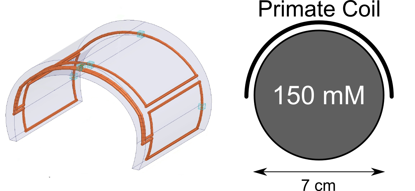
MRI acquisitions were performed on our 7T Magnetom MRI (Siemens, Erlangen, Germany) with a home-made transceiver 23Na radiofrequency coil (Fig. 1) and a cylindrical phantom (diameter 7cm, 150mM NaCl). A 3D gradient echo sequence was used to acquire 45 different radiofrequency spoiling increments sequentially, leading to a total acquisition time of 5h16min (TR/TE=20ms/3.2ms, Bw: 220Hz/px, resolution: 6mm3 isotropic, FoV: 192x192x160mm3). Spoiling gradients were restricted to the readout direction, equal to 22 times the readout gradient momentum. The data were fitted voxel-wised to Bloch-Torrey equations to estimate M0, R1, R2, ADC, and the flip angle maps using Matlab (The MathWorks, Natick, USA).Results and Discussion
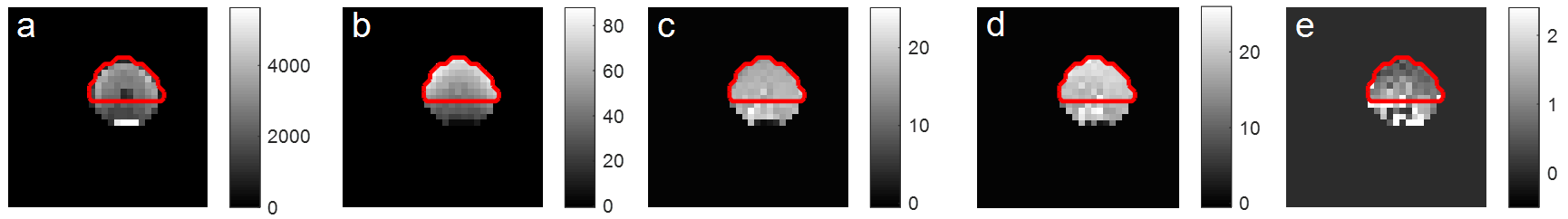
Given the hemi-cylindrical geometry of the coil (Fig.1.), a region-of-interest (ROI) was defined over the top of the phantom as shown in Fig.2. In this ROI, mean R1 was 17.3±1.43s-1 (T1=58ms), R2 19.9±2.13s-1 (T2=50ms), ADC 1.11±0.50x10⁻⁹m².s-1 and flip angle 51.5±10.9° (Fig. 2).
For saline solutions of 140-150mM mimicking the physiological CSF, T1 values of 50-55ms and T2 values of 55-65ms are reported in the literature7,8 for in vivo conditions. Diffusion coefficient were estimated for sodium in the rat brain at 25°C to be 1.15x10⁻⁹m².s-1 9 and in sodium fluorine in aqueous solution at 25°C to be 1.3x10⁻⁹m².s-1 10. Overall, our results were in good agreement with these data, with an error of 5% for T1, 9% for T2 and 8% for ADC.
Conclusion
In this preliminary in vitro study, we have demonstrated the possibility to use the QuICS method, not only to estimate accurately the T1, T2, FA, M0 and ADC simultaneously for 23Na at physiological concentration at UHF, but to acquire 3D maps for all of them. This is particularly exciting considering the difficulty of conventional approaches to estimate parameters such as the ADC for nuclei with short T2 relaxation times. To the knowledge of the authors, this is the first time that such multi-parametric extraction is reported in the context of X-nuclei imaging.
Diffusion was the most complicated parameter to estimate and required the full range of RF spoiling increments described above. However, the estimation of M0, R1 and R2 was achieved with good precision in less than an hour. For now, the long acquisition time remains a significant hurdle to translate this method to clinical or preclinical MRI. We are aiming at accelerating our method by using shorter TE, non-Cartesian sampling trajectories11, a better coil configuration12 and eventually less FA, RF and gradient spoiling steps. An optimization algorithm based on Fisher information and Cramér-Rao lower bound will be used for this purpose13,14.
Acknowledgements
This project received financial support from France Life Imaging ("qMRI" starter project).References
1. D. Ma et al., “Magnetic resonance fingerprinting,” Nature, vol. 495, no. 7440, pp. 187–192, Mar. 2013.
2. J. b. m. Warntjes et al., “Novel method for rapid, simultaneous T1, T*2, and proton density quantification,” Magn. Reson. Med., vol. 57, no. 3, pp. 528–537, Mar. 2007.
3. J. b. m. Warntjes, et al., “Rapid magnetic resonance quantification on the brain: Optimization for clinical usage,” Magn. Reson. Med., vol. 60, no. 2, pp. 320–329, Aug. 2008.
4. R. Heule et al., “Triple echo steady-state (TESS) relaxometry,” Magn. Reson. Med., vol. 71, no. 1, pp. 230–237, Jan. 2014.
5. P. Schmitt et al., “Inversion recovery TrueFISP: Quantification of T1, T2, and spin density,” Magn. Reson. Med., vol. 51, no. 4, pp. 661–667, Apr. 2004.
6. L. de Rochefort, “Encoding with Radiofrequency Spoiling, Equilibrium States and Inverse Problem for Parametric Mapping,” Proc. Intl. Soc. Mag. Reson. Med., 2015, p. 445.
7. G. Madelin and R. R. Regatte, “Biomedical applications of sodium MRI in vivo,” J. Magn. Reson. Imaging, vol. 38, no. 3, pp. 511–529, Sep. 2013.
8. N. J. Shah et al., “Imaging of sodium in the brain: a brief review,” NMR Biomed., vol. 29, no. 2, pp. 162–174, Feb. 2016.
9. J. A. Goodman, C. D. Kroenke, G. L. Bretthorst, J. J. H. Ackerman, and J. J. Neil, “Sodium ion apparent diffusion coefficient in living rat brain,” Magn. Reson. Med., vol. 53, no. 5, pp. 1040–1045, May 2005.
10. A. C. F. Ribeiro et al., “Diffusion Coefficients of Sodium Fluoride in Aqueous Solutions at 298.15 K and 310.15 K,” ResearchGate, vol. 57, no. 2, pp. 410–4, Jun. 2010.
11. A. Coste et al., “Assessment of benefit to use a non-Cartesian trajectory and nonlinear reconstruction method compared to a Cartesian strategy for fast 31P MRI,” Proc. Intl. Soc. Mag. Reson. Med. 24, 2016, p. 3940.
12. G. Shajan et al., “Three-layered radio frequency coil arrangement for sodium MRI of the human brain at 9.4 Tesla,” Magn. Reson. Med., vol. 75, no. 2, pp. 906–916, Feb. 2016.
13. L. de Rochefort et al., “In Vivo Feasibility of Multi-Parametric Mapping Based on Fast Steady-State Sequences,” Proc. Intl. Soc. Mag. Reson. Med., 2016, p. 1823.
14. R. Valabrègue and L. de Rochefort, “Fisher Information Matrix for Optimizing the Acquisition Parameters in Multi-Parametric Mapping Based on Fast Steady-State Sequences,” Proc. Intl. Soc. Mag. Reson. Med., 2016, p. 1569.
Figures