5624
Design and construction of a novel double-tuned 1H/19F coil using PIN-diode switches at 9.4T1Institute of Neuroscience and Medicine-4, Forschungszentrum Juelich, Juelich, Germany, 2Faculty of Medicine, Department of Neurology, JARA, RWTH Aachen University, Aachen, Germany
Synopsis
A double-tuned 1H/19F coil using PIN-diode switches was developed and evaluated its performance on a 9.4T preclinical MRI scanner. This proposed design uses an inductor rather than a capacitor in series with the PIN-diode so that the resonance frequency is shifted in the opposite direction compared to the conventional method. This is a key difference from the previous developments and in this way we can maintain the SNR or image quality of the X-nuclei (therefore 19F); the SNR is nearly as good as a single-tuned 19F coil.
PURPOSE
A Flurorine-19 (19F) based MR technique as a molecular imaging tool allows one to directly detect and to quantify extremely specific cells1,2. Double-tuned coils are often used to detect the exogeneous 19F substance and its metabolites without contamination from endogeneous background signals. Since the 1H and 19F nucleus resonate at frequencies that are close to each other (gyromagnetic ratio of 1H/19F = 42.58/40.05 MHz/Tesla), the conventional technique of using traps is not easily implementable. There are several approaches to design 1H/19F coils but they also have limitations in the signal-to-noise ratio (SNR)3,4. PIN-diodes as an electronic switch can be an excellent candidate to design a double-tuned coil for this purpose. Coils with PIN-diodes, in general, are likely to be used together with capacitors in series in order to generate double-resonance frequencies5,6. However, the sensitivity of the non-proton coil degrades due to the resistance of the PIN-diodes especially when forward bias is applied. On the other hand, when a reverse voltage is applied, loss due to the PIN-diodes can be negligible – normally used for 1H. This is problematic because sensitivity in X-nuclei is already insufficient compared to 1H and additional loss is not desirable. In this work, we have designed and constructed a double-tuned 1H/19F coil using PIN-diode switches and evaluated its performance on a 9.4T preclinical MRI scanner. This proposed design uses an inductor rather than a capacitor in series with the PIN-diode so that the resonance frequency is shifted in the opposite direction compared to the conventional method. This is a key difference from the previous developments and in this way we can maintain the SNR or image quality of the X-nuclei (therefore 19F); the SNR is nearly as good as a single-tuned 19F coil.METHODS
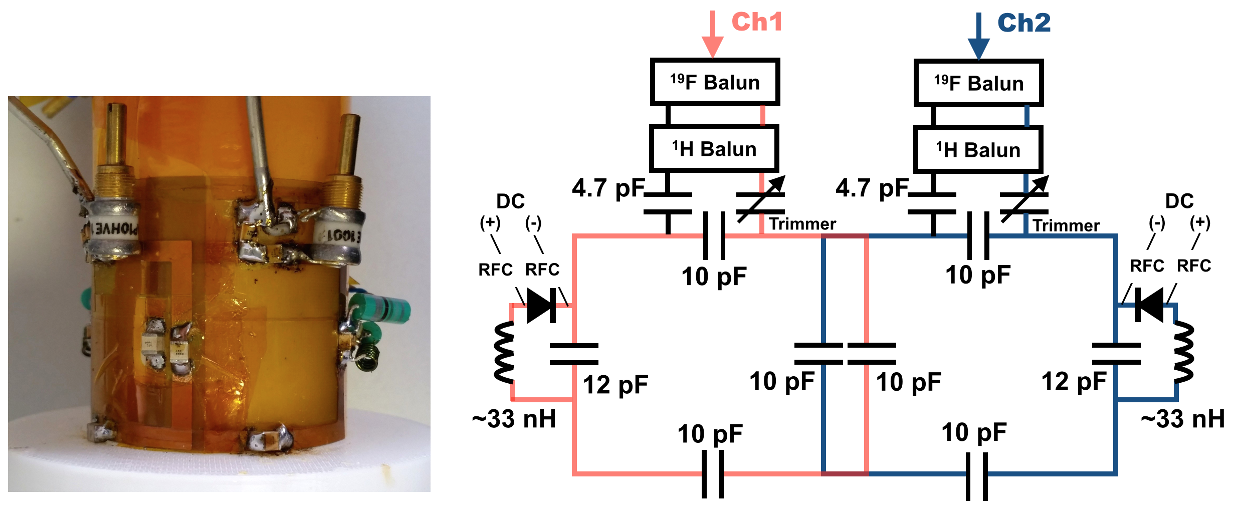
As shown in Fig. 1, the double-tuned coil was designed as overlapped surface loops, which operates in a quadrature mode. Each loop of the coil contains a PIN-diode switch in series with an inductor but in parallel to one of the capacitors on the loop. The double-resonant coil was adjusted to 19F when the reverse bias was applied (PIN-diode OFF, -30V), while it was switched to 1H when forward current was flown (PIN-diode ON, 100 mA). A home-built active transmit/receive switch incorporated in a Wilkinson unit was used operating for both frequencies. The response of the double-tuned coil was evaluated and compared with 1H and 19F single-tuned coils using a network analyser on the bench. Imaging experiments using a phantom containing 150 mM NaF were carried out on a 9.4T preclinical scanner.RESULTS
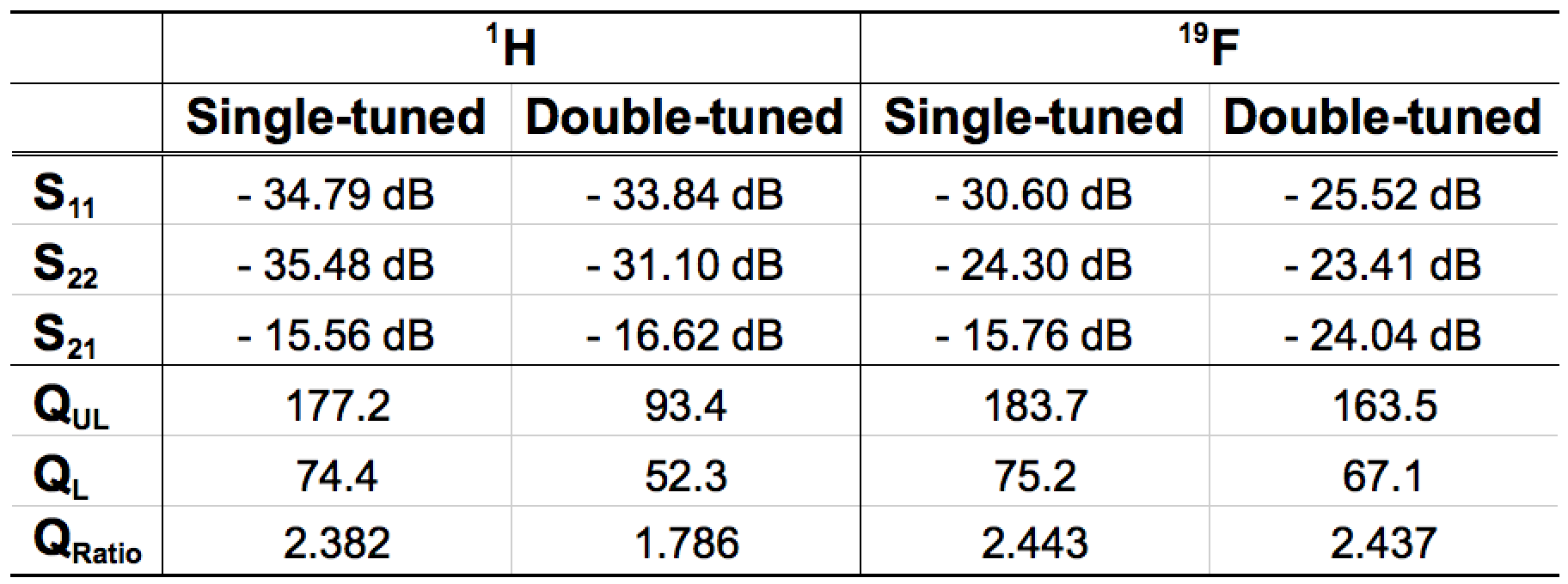
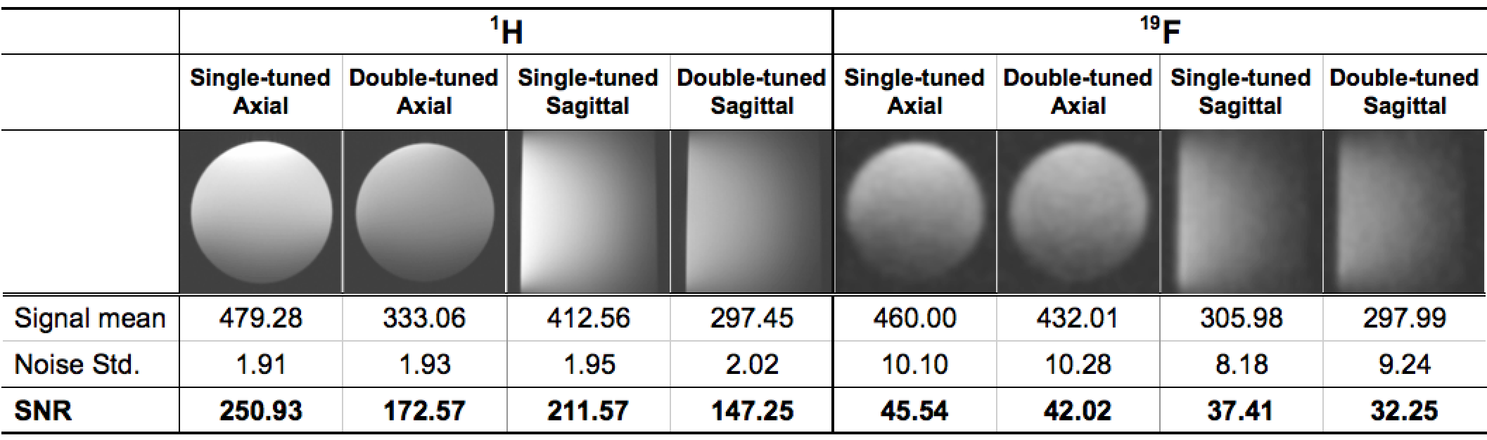
Table 1 shows S-parameters and Q-factors of single- and double-tuned coils measured and compared with/without the phantom. All the coils were well tuned at resonance frequencies corresponding to 1H (400 MHz) or 19F (376 MHz) and matched well to 50 ohm (<-25 dB). Fig. 2 shows the axial and sagittal images obtained using the scan parameters for 1H: TR = 100 ms, TE = 4.69 ms, 2 averages, 200 um x 200 um x 1 mm and scan time ~ 38 seconds. The scan parameters for 19F were: TR = 450 ms, TE = 2.48 ms, 32 averages, 1 x 1 x 1 mm and scan time ~ 10:06 minutes. Fig. 2 also shows SNR values calculated from the signal mean value divided by the standard deviation of the noise image (Tx = 0V) in the region where a rat brain would be located.DISCUSSION/CONCLUSIONS
We have built a PIN-diode switchable double-tuned RF coil and have demonstrated the performance of the coil. The SNR of 19F obtained using this proposed design is shown to be nearly as good as that of a single-tuned coil, i.e., approximately 90%. It may also be possible to improve further this figure by carefully selecting RF chokes and by improving cabling and ground. A similar concept, used for different purposes, has been implemented into most detune circuits on multi-channel Rx-only 1H coils7,8.Acknowledgements
No acknowledgement found.References
1. Srinivas M, Heerschap A, Ahrens ET, et al. 19F MRI for quantitative in vivo cell tracking. Trends Biotechnol. 2010;28:363-370.
2. Ruiz-Cabello J, Barnett BP, Bottomley PA, Bulte JWM. Fluorine (19F) MRS and MRI in biomedicine. NMR Biomed. 2011;24:114-129.
3. Gajawada G, Li T, Couch MJ, Fox MS, Albert M. A 19F – 1H linear dual tuned RF birdcage coil for rat lung imaging at 3T. ISMRM. 2015;1461.
4. Bowyer P, Finnigan J, Marsden B, Taber B, Zens A. Using magnetic coupling to implement 1H, 19F, 13C experiments in routine high resolution NMR probes. J. Magn. Reson. 2015;261:190-198.
5. Lim H, Thind K, Martinez-Santiesteban FM, Scholl TJ. Construction and Evaluation of a Switch-Tuned 13C - 1H Birdcage Radiofrequency Coil for Imaging the Metabolism of Hyperpolarized 13C-Enriched Compounds. J. Magn. Reson. Imaging. 2014;40:1082-1090.
6. Lee S-P, Choi I-Y, Kim S-G. Rapidly switchable RF coil for 19F/1H NMR studies. ISMRM. 2000;1416.
7. Fujita H, Zheng T, Yang X, Finnerty MJ, Handa S. RF surface receive array coils: the art of an LC circuit. J. Magn. Reson. Imaging. 2013;38:12-25.
8. van de Bank BL, Orzada S, Smits F, et al. Optimized 31P MRS in the human brain at 7 T with a dedicated RF coil setup. NMR Biomed. 2015;28:1570–1578.
Figures