5613
Optimization of Regularization Parameters of Compressed Sensing Reconstruction for Fast Phosphorus MR Spectroscopic Imaging of Human Brain.1Institute of Biomedical Engineering, Bogazici University, Istanbul, Turkey, 2Advanced Diagnostic Imaging, Philips Healthcare, Best, Netherlands
Synopsis
This study aims at investigating the effects of compressed sensing data acquisition and reconstruction factors for accelerated phosphorus MR spectroscopic imaging (31P-MRSI). Simulated 31P MRSI datasets containing healthy and tumor regions were created based on the metabolite information of brain tumor patient 31P-MRSI acquired at 3T. k-space data were randomly undersampled with three different reduction factors while preserving the central portion for different noise levels, reduced datasets were reconstructed using compressed sensing by combining eleven different total variation and L1-norm penalties. Findings showed that data acquisition pattern and reconstruction parameters have a significant effect on the resultant 31P-MRSI spectral quality.
Purpose
Phosphorus MR spectroscopic imaging (31P-MRSI) provides important information about energy metabolism, oxygen state and pH for brain tumors. However, MR sensitivity of phosphorus is 15 times less than that of proton and it also has a lower gyromagnetic ratio (17.24 MHz/T), so larger voxels and averaging several acquisitions are necessary for adequate signal to noise ratio (SNR) for 31P-MRSI. Previous studies have reported the feasibility of compressed sensing1 to accelerate 31P-MRSI with low SNR penalty2. In this study, we investigate the impact of factors governing compressed sensing data acquisition and reconstruction, such as the undersampling pattern, matrix size, noise and the regularization parameters on the performance of accelerated 31P-MRSI.Methods
Phosphorus MR spectra of seven patients diagnosed with brain tumors (2 Non-Hodgkin’s lymphoma (NHL), 1 grade I oligodendroglioma, 1 metastasis, 1 grade II oligodendroglioma, 2 grade II astrocytoma, average age=47.3±12.8 years) that were scanned on a clinical Philips 3T scanner were retrospectively quantified to estimate the metabolite peak level differences of healthy brain and brain tumor3. The amplitudes of phosphocreatine (PCr), glycerophosphorylcholine (GPC), glycerophosphorylethanolamine (GPE), inorganic phosphate (Pi), phosphorylcholine (PC), phosphorylethanolamine (PE), γ-adenosinetriphosphate (ATP), α-ATP and β-ATP peaks of the brain tumor spectrum were set to be 0.59, 0.93, 0.75, 0.62, 0.82, 0.81, 0.75, 0.72 and 0.72 times the peak amplitudes of the simulated healthy brain spectrum, respectively. Two dimensional 8x8, 16x16 and 32x32 31P-MRSI datasets containing a tumor voxel at the corner of top left region, and healthy spectra at the rest of the array were simulated in MATLAB (The Mathworks Inc., Natick, MA). Four noise levels that had a standard deviation equal to the 8%, 30%, 40% and 50% of the maximum signal intensity of the healthy signal were added to the spectra. Six different random undersampling patterns that have total reduction factors of 4, 5 and 10, which fully samples a high (6.25% of the total array size) or low (1.56% for 16x16, 32x32 and 4.69% for 8x8) portion of the k-space center, were generated for each matrix size. k-space data were undersampled with these patterns. Each dataset was reconstructed by using a 3D compressed sensing reconstruction algorithm2 modified from the SparseMRI package1 with a combination of eleven different total variation (α) and L1-norm (λ) penalties. Root mean square error (RMSE) of each spectra was calculated.
Results
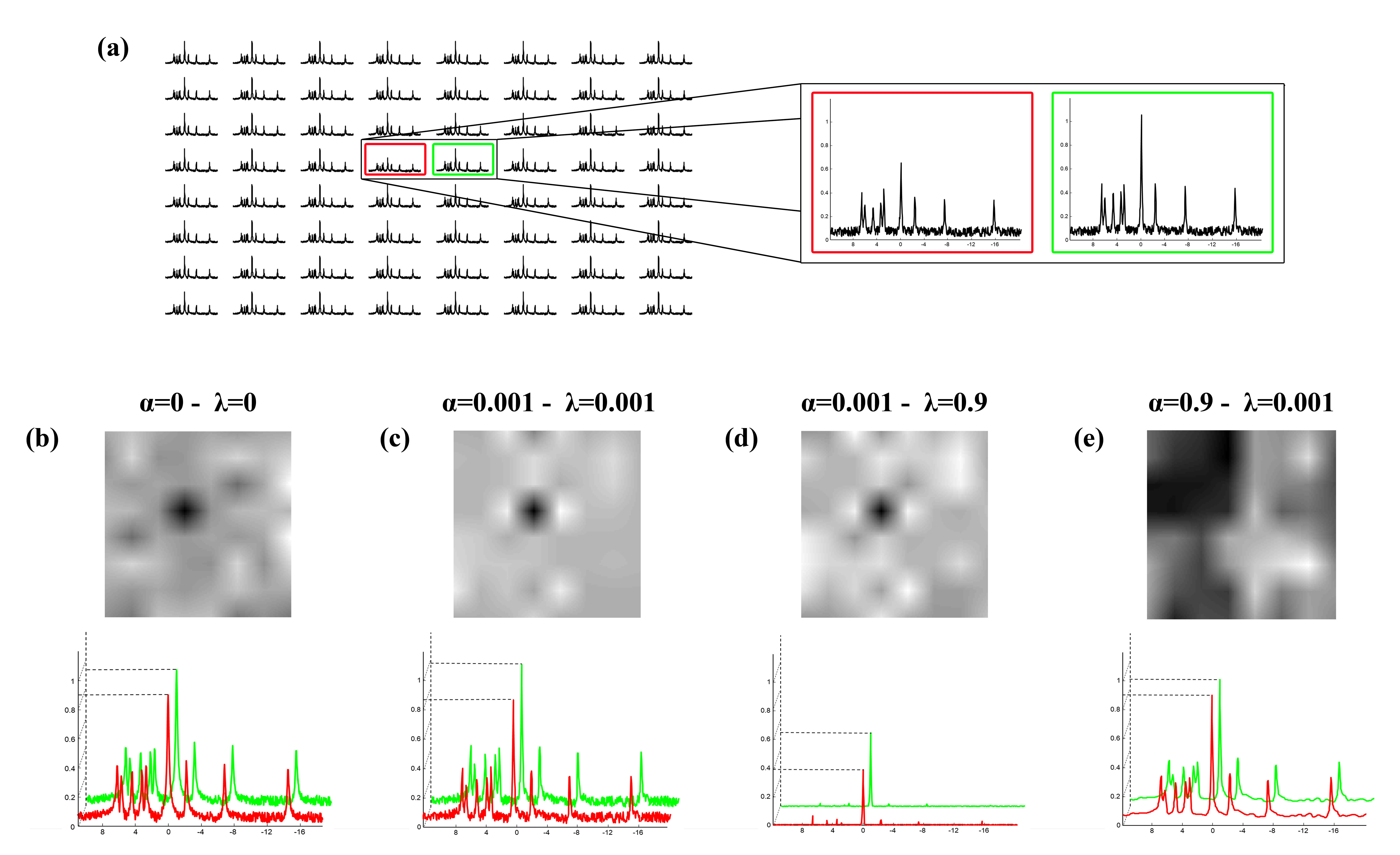
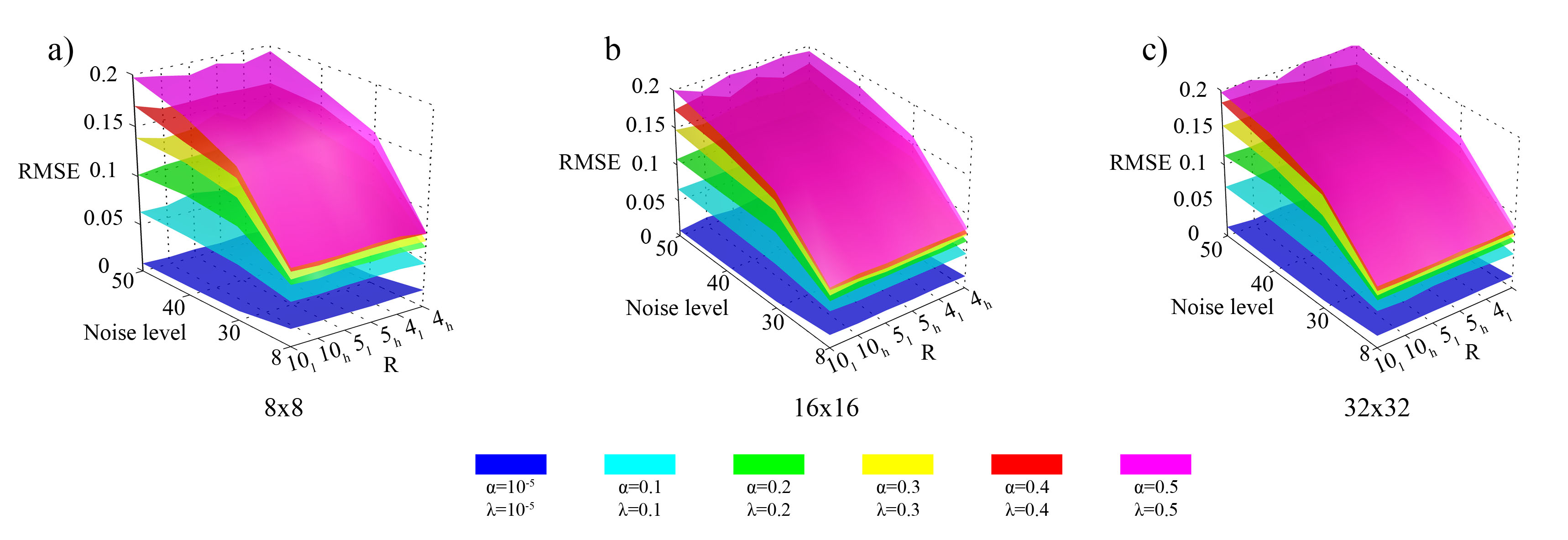
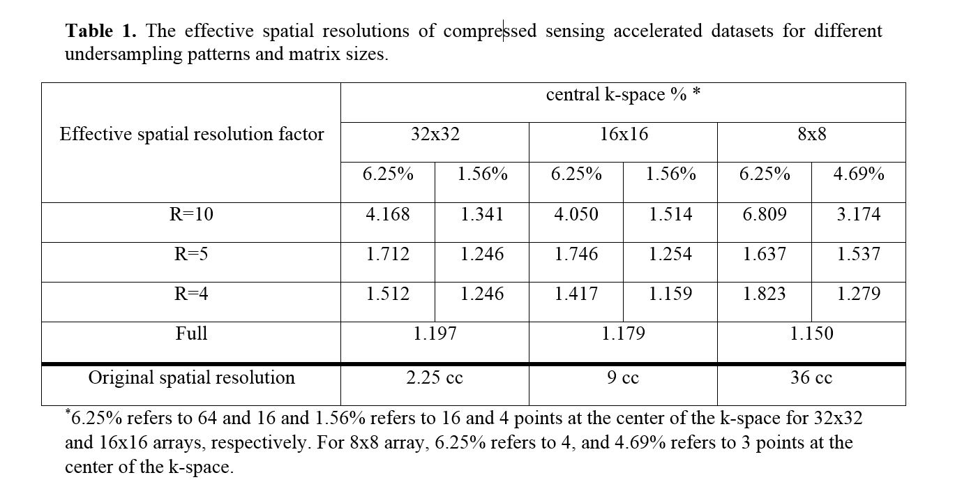
The effective spatial resolution factor increased with reduction factor and sampling more at the k-space center for each matrix size (Table 1).The exclusion of L1-norm and total variation penalties from the inverse problem resulted in a less distinguishable tumor and healthy spectra (Figure 1b) than using a low value of 0.001 for both penalties (Figure 1c). Increasing the λ penalty resulted in a baseline removal of the spectra and loss of peak intensities (Figure 1d), and increasing the α penalty resulted in a smearing of the tumor voxel intensity on the PCr frequency image and the loss of the tumor region contrast (Figure 1e). The results indicated that using lower values for both penalties resulted in a narrow point spread function and a more accurate definition of the tumor voxel location based on its spectrum. Figure 2 shows the RMSE values of the compressed sensing reconstructed datasets with six different k-space undersampling patterns and four different noise levels, for 8x8, 16x16 and 32x32 matrix sizes. The RMSE values increased with the noise level for all the datasets. On the other hand, slightly less RMSE values were observed in high k-space central sampling patterns for all reduction factors. Matrix sizes did not highly affect the RMSE values. Also it was observed that higher L1-norm penalties resulted in higher RMSE values due to the denoising effect.Discussion
Phosphorus MR spectroscopic imaging provides valuable information about brain tumors, but 31P-MRSI has acquisition time and signal intensity limitations that prevent the widespread usage in clinical settings. Compressed sensing reconstruction enables shorter acquisition time and enhances the signal to noise ratio. The center of the k-space holds the high signal intensities, so sampling more at the central k-space resulted in better spectral quality, and lower RMSE values than sampling a lower portion of the central k-space. The simulation results showed that total variation and L1-norm penalties affected the definition of tumor location.Conclusion
The results of this study showed that the signal quality of resultant 31P-MRSI was related with the choice of the undersampling pattern, noise level and regularization parameters of compressed sensing. Compressed sensing accelerated 31P-MRSI could be used in the clinical settings for imaging brain tumors with the appropriate set of the undersampling and reconstruction parameters.Acknowledgements
This study was supported by TUBITAK Career Development Grant 112E036, EU Marie Curie IRG grant 256528, and Philips Healthcare through a research agreement.References
1. Lustig, M., D. Donoho, and J.M. Pauly, Sparse MRI: The application of compressed sensing for rapid MR imaging. Magn Reson Med, 2007. 58(6): p. 1182-95.
2. Hatay, G., et al. Comparison of 2D Iterative Frame Based and 3D Direct Compressed Sensing Reconstruction for Accelerated Phosphorus MR Spectroscopic Imaging of Human Brain. in In Proceedings of the 22nd Annual Meeting of ISMRM. 2014. Milan, Italy.
3. Citak Er, F., et al., Classification of Phosphorus Magnetic Resonance Spectroscopic Imaging of Brain Tumors Using Support Vector Machine and Logistic Regression at 3T. Conf Proc IEEE Eng Med Biol Soc, 2014. 2014: p. 2392-5.
Figures