5586
A MatLab-based simulation program (tcaSIM2) for predicting NMR spectra and MS data for 13C tracer experiments1Advanced Imaging Research Center, University of Texas Southwestern Medical Center, Dallas, TX, United States, 2Chemistry, University of Texas at Dallas, Richardson, TX, United States, 3Advanced Imaging Research Center, UT Southwestern Medical Center
Synopsis
A MatLab-based program is presented for predicting 13C NMR spectra and mass 13C isotopomer data of various tissue metabolites in a 13C tracer experiments. The program is useful for predicting changes in 13C multiplet patterns in NMR spectra and changes in mass isotopomer ratios in mass spectral data as a tissue responds to changes in flux of various substrates through completing pathways involving mitochondrial metabolism. The program tcaSIM2 (copies available free of charge) is also valuable for teaching metabolism and analysis of 13C NMR data and mass spec data in metabolic tracer experiments.
Introduction:
The use of stable isotopes for tracing metabolism in cells, perfused tissues, and in vivo has grown substantially over the past few years. We have written extensively on analysis of 13C NMR spectra collected at steady-state for providing quantitative flux data (13C isotopomer analysis).1 Ultra-sensitive cryo-probes combined with high field magnets now allow 13C NMR spectra to be collected on samples approaching the size of a typical tissue biopsy. For example, 13C NMR analysis of surgical specimens collected from pre-operative cancer patients after infusion of various 13C-enriched substrates has revealed novel oncogenic metabolic pathways.3 Other approaches, such as hyperpolarized (HP) 13C metabolic imaging, capture in vivo metabolic characteristics of specific organs or diseased tissue. Dynamic HP flux data are difficult to validate, but adding an isotopomer analysis to an HP experiment is one approach.4 In both cases, the costly nature of the experiments demand thoughtful consideration of tracer design and expected isotopomer outcomes. Here, we introduce a MatLab-based simulation program called tcaSIM2 that allows one to predict both dynamic and steady-state NMR and MS data for complex metabolic systems.Methods:
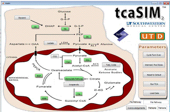
A C++ program originally written to run in DOS was rewritten and compiled in MatLab for use on any computer platform. The interface shown in Figure 1 allows the operator to select 13C-labeling patterns for glycerol, lactate, fatty acids or CO2 (for possible labeling via a carboxylation pathway) and to choose relative activities of PDH (pyruvate dehydrogenase), yPC (pyruvate carboxylase), TPI (triose phosphate isomerase), GK (glycerol kinase), and PK (pyruvate kinase). The output provides calculated 13C NMR spectra for all 3-5 carbon TCA cycle intermediates, alanine and lactate, and glucose for those tissues undergoing active gluconeogenesis.Results:
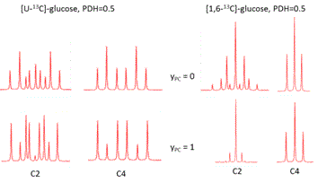
An example of how tcaSIM2 can be helpful in planning a 13C metabolic experiment. Consider an experimental plan to infuse 13C-enriched glucose into a glioma patient prior to surgery. A 13C NMR spectrum of the tumor extract removed during surgery could be used to evaluate the contribution of glucose to energy production and the extent of anaplerosis in tumor metabolism. One might initially ask, should I choose [1,6-13C]glucose or [U-13C]glucose for the experiment? The simulated 13C NMR spectra of glutamate ± flux of pyruvate into the TCA cycle via yPC (anaplerosis) are shown in Figure 2. These spectra show that the glutamate spectrum is sensitive to influx of labeled pyruvate into the TCA cycle via pyruvate carboxylase regardless of which labeled glucose is chosen for the experiment. However, they also demonstrate that the glutamate C2 resonance is quite sensitive to anaplerosis while the glutamate C4 resonance is less sensitive when [1,6-13C]glucose is chosen for the experiment. These results suggests that [U-13C]glucose may be a better choice for the experiment.
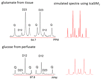
Comparison of experimental NMR spectra with those predicted by tcaSIM2. Figure 3 illustrates experimental 13C NMR data from a liver perfused with a cocktail of unlabeled substrates plus [U-13C]propionate with a goal of evaluating gluconeogenesis (GNG), total anaplerosis (yS plus yPC), and pyruvate cycling.2 A 13C isotopomer analysis of the multiplets appearing in the 13C NMR spectrum of glutamate isolated from liver and glucose isolated from perfusate indicated that total anaplerosis was 6-fold higher than TCA cycle flux, GNG was 4-fold higher that TCA cycle flux, and surprisingly, pyruvate cycling flux was 2-fold greater than TCA cycle flux. The spectra of glutamate and glucose predicted by tcaSIM2 using these flux parameters are shown next to the experimental spectra in Figure 3. The agreement is obvious.
Conclusions:
A second generation MatLab-based program, tcaSIM2, is quite valuable for predicting 13C NMR spectra and mass isotopomer data for a variety of metabolites in tracer experiments. We have found the program to be extremely useful in teaching metabolism and tracer technologies to students and colleagues interested in 13C tracer experiments in animals and human subjects.Acknowledgements
Continued development of tcaSIM2 over many years has been supported by grants from the National Institutes of Health (P41-EB015908 to CRM and R37-HL034557 to ADS).References
1. (a) Malloy, C. R.; Sherry, A. D.; Jeffrey, F. M., Evaluation of carbon flux and substrate selection through alternate pathways involving the citric acid cycle of the heart by 13C NMR spectroscopy. Journal of Biological Chemistry 1988, 263 (15), 6964-6971; (b) Malloy, C. R.; Sherry, A. D.; Jeffrey, F. M., Analysis of tricarboxylic acid cycle of the heart using 13C isotope isomers. American Journal of Physiology 1990, 259 (3), H987-H995; (c) Sherry, A. D.; Jeffrey, F. M. H.; Malloy, C. R., Analytical solutions for 13C isotopomer analysis of complex metabolic conditions: substrate oxidation, multiple pyruvate cycles, and gluconeogenesis. Metabolic Engineering 2004, 6 (1), 12-24.
2. Burgess, S. C.; Hausler, N.; Merritt, M.; Jeffrey, F. M. H.; Storey, C.; Milde, A.; Koshy, S.; Lindner, J.; Magnuson, M. A.; Malloy, C. R.; Sherry, A. D., Impaired Tricarboxylic Acid Cycle Activity in Mouse Livers Lacking Cytosolic Phosphoenolpyruvate Carboxykinase. Journal of Biological Chemistry 2004, 279 (47), 48941-48949.
3. Maher, E. A.; Marin-Valencia, I.; Bachoo, R. M.; Mashimo, T.; Raisanen, J.; Hatanpaa, K. J.; Jindal, A.; Jeffrey, F. M.; Choi, C.; Madden, C.; Mathews, D.; Pascual, J. M.; Mickey, B. E.; Malloy, C. R.; DeBerardinis, R. J., Metabolism of [U-13C]glucose in Human Brain Tumors In Vivo. NMR in Biomedicine 2012, 25 (11), 1234-1244.
4. Yang, C.; Harrison, C.; Jin, E. S.; Chuang, D. T.; Sherry, A. D.; Malloy, C. R.; Merritt, M. E.; DeBerardinis, R. J., Simultaneous Steady-state and Dynamic 13C NMR Can Differentiate Alternative Routes of Pyruvate Metabolism in Living Cancer Cells. Journal of Biological Chemistry 2014, 289 (9), 6212-6224.
Figures