5583
Graphene for MRI Applications at 7T: Opportunities for SAR reduction1Imago7, Pisa, Italy, 2IRCCS Stella Maris Foundation, Pisa, Italy, 3Xidian University, Xidian, People's Republic of China, 4University of Central Florida in Orlando, Orlando, FL
Synopsis
SAR management is critical at ultra-high field (UHF) strength where RF field energy deposition in the subject increases and its distribution becomes very inhomogeneous. Here we illustrate simulation results for a test example in order to show how a graphene sheet can be used to obtain a SAR reduction without sacrificing the coil efficiency significantly. Specifically, the presence of the graphene sheet leads a maximum local SAR reduction up to 47%. Thus, from the simulation here shown, it follows that graphene sheets can be successfully used in MRI applications at 7T for enhancing the safety with respect to SAR issue
Target audience.
RF Engineers interested in SAR aspects at UHF.Purpose.
SAR management is critical at ultra-high field (UHF) strength where RF field energy deposition in the subject increases and its distribution becomes very inhomogeneous and subject-dependent1. Here we illustrate simulation results for a test example in order to show how a graphene sheet can be used to obtain a SAR reduction without sacrificing the coil efficiency significantly.Methods.
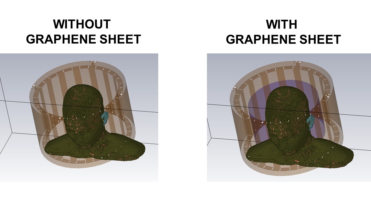
Electromagnetic Simulations. We used the software HFSS, ANSYS, to simulate a volume coil loaded by a human head. Specifically, we simulated a transmit-receive shielded 16 elements 1H high-pass birdcage head coil manufactured by Nova Medical (Wilmington, MA, USA), operating in quadrature at 298 MHz. The elements (copper flat strips having width of 2.5 cm and thickness of 35 μm) of the coil are placed equally spaced along a circle of diameter of 29.5 cm; the elements are connected through two copper end-rings (having width of 1 cm). The diameter of the copper shield is 37.5 cm while the length is 27.5 cm. The coil was loaded by a homogeneous human head extracted from an anatomic adult human model (derived from SAM) as shown in Figure 1a. Quadrature feeding has been employed using 4 sources, equally displaced by π/2 azimuthally, with a relative electrical phase shift of π/2. The coil has been tuned to the frequency of 298 MHz and matched using a capacitive matching circuit1, achieving Sxx<-10 dB and Sxy<-10 dB. RF fields and SAR inside the head were calculated when applying 1 W of input power at each source. The average B1+ magnitude calculated in the axial slice crossing the eyes was computed, together with maximum local SAR (10 g, continuous RF wave excitation) calculated in the entire head.
Graphene Characterization and Modelling. Graphene has recently attracted tremendous interest in various research areas due to its exceptional electrical and mechanical properties 2. It is now commonly used in techniques for reducing the SAR in mobile devices 3. Explanation on the unique band physics of graphene is but can be found in the plethora of references on the topic 4. For our purpose, it is sufficient here to briefly discuss the main quantity of interest for graphene in electromagnetic (EM) applications. Since graphene is mono-atomic layer, it is perfectly modeled by its surface conductivity. This surface conductivity depends on graphene unique band structure and on a number of parameters including temperature, scattering rate, fermi energy, electron velocity, pre-doping, frequency, as well as potential bias. At microwave, the surface impedance of graphene sheet has a real part of 10Ω÷100 Ω and an imaginary part of -0.001Ω ÷ -0.1Ω. Here, we considered the three following cases: i) 85.07-j0.016Ω; ii) 56.71-j0.01Ω; iii) 42.53-j0.007Ω For each one of the three cases, we repeated the electromagnetic simulations introducing a graphene sheet between the coil and the head, as shown in Figure 1b. The graphene sheet has height of 15 cm and diameter of 25.2 cm The graphene sheet is modelled as impedance surface, and it is included in simulations using the procedure shown in 3.
Results.
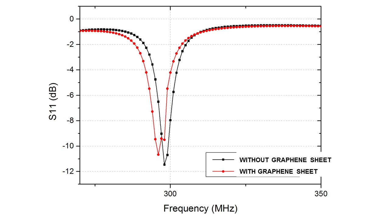
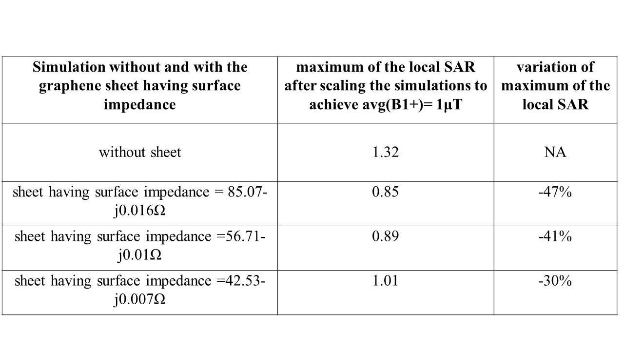
Figure 2 shows the s11 of volume coil loaded by a human head without and with the graphene sheet having 85.07-j0.016Ω. Table 1 summarizes the results obtained by applying simulation to the human head without and with the three different graphene sheets. Specifically, the second column shows the maximum of the local SAR after scaling the simulations to achieve the B1+ slice average value of 1μT (calculated in the axial slice crossing the eyes). The third column shows the variation of maximum of the local SAR (after scaling the simulations to achieve the B1+ slice average value of 1μT) with respect to human head without graphene sheet.Discussions and Conclusion.
The presence of the graphene sheet leads to a slight detuning of the coils; this turns out also on a lost of B1+ efficiency, here estimated in less than 10%. Such lost in B1+ efficiency can be easily compensated during transmission. Concerning the SAR, the presence of the graphene sheet leads a maximum local SAR reduction up to 47%. Thus, from the simulation here shown, it follows that graphene sheets can be successfully used in MRI applications at 7T for enhancing the safety with respect to SAR issueAcknowledgements
No acknowledgement found.References
1. Tiberi G, Fontana N, Costagli M, et al. Investigation of maximum local specific absorption rate in 7T magnetic resonance with respect to load size by use of electromagnetic simulations, Bioelectromagnetics, 2015, 36(5):358-366.
2. Perruisseau-Carrier J, Graphene for Antenna Applications: Opportunities and Challenges from Microwaves to THz, Loughborough Antennas & Propagation Conference, 2012, UK.
3. Pelletti C, L Li, M Abdel-Mageed, et al, Techniques for reducing the SAR in mobile devices by using graphene-type absorbing materials, 9th European Conference on Antennas and Propagation (EuCAP), 2015: 1-3.
4. Geim GK, Novoselov KS, The rise of graphene, Nature Matter, 2007, 6:183-191
Figures