5578
The effect of variable amniotic fluid conductivity and fetal tissues properties on B1+ and local SAR for fetal imaging at 3T1Division of Imaging Sciences & Biomedical Engineering, King's College London, London, United Kingdom
Synopsis
Effects on B1+ and local specific absorption rate of varying the conductivity of fetal tissues and amniotic fluid ($$$\sigma_{AF}$$$) in a model of a 7 month pregnant woman within a 3T birdcage coil are investigated numerically. Results indicate that a realistic value of $$$\sigma_{AF}$$$ is required to estimate power to produce a chosen B1+ in the fetus and fetal SAR10g. Fetal properties adjusted for gestational age impact on B1+ and result in increased fetal SAR10g compared to adult value based simulations. Smaller changes in SAR10g are predicted between detailed fetal models and homogeneous ones with volume weighted average dielectric properties.
Purpose
There is some uncertainty regarding dielectric properties of both fetal tissues, which differ from adult equivalents by larger water content that changes throughout gestation, and amniotic fluid (AF) whose composition can change over time. In this study we examine the effect of varying the conductivity of the fetal tissues and AF, and the position of the fetus in a model of a 7 month pregnant woman within a 3T birdcage coil on RF magnetic field (B1+) and local specific absorption rate averaged over 10g of tissue (SAR10g).Methods
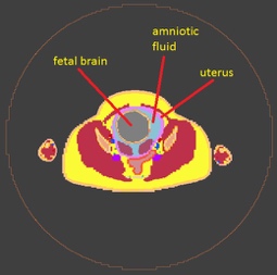
The pregnant model1 was positioned fetal brain centred (Figure 1) within a 60 cm diameter, 40 cm long band-pass shielded coil which was excited from two ports in quadrature. RF fields and SAR10g were calculated using a FDTD solver (Sim4Life, Zurich Med Tech). Maternal tissue properties were taken from a standard database2. Amniotic fluid conductivity ($$$\sigma_{AF}$$$) has been measured at ~1.25 S/m at 127 MHz 3 and shows little change up to ~30 weeks gestation; it falls to ~1.1 S/m at full term 4 Data from fetal animal tissues5 and predictions based on mixture equations6,7 suggest that the conductivity of some fetal tissues may be comparable to or greater than $$$\sigma_{AF}$$$ 7,8.
Simulations were performed for a range of fetal tissue dielectric properties determined using (a) the approach reported by Wang et al 6 (Model A), adult human values 2 (Model B), and a homogeneous fetus with volume weighted mean dielectric properties of model A (model C). The effects of increasing $$$\sigma_{AF}$$$ to 2.14 S/m (that of cerebrospinal fluid ($$$\sigma_{CSF}$$$ )) or decreasing it to 0.72 S/m (that of adult muscle tissue)) and moving the fetus within the uterus were also investigated. The resulting B1+ fields were evaluated by averaging over the fetus or uterus and axially from z=-25 mm to +25 mm).
Results
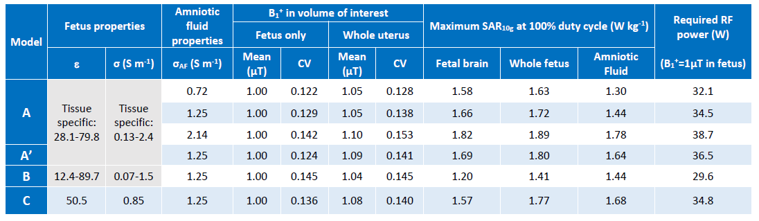
The values of B1+ and SAR10g normalised so that the mean B1+ = 1 $$$\mu T$$$ in the fetus within the axial volume of interest, are shown in Table 1. The SAR10g values assume 100% duty cycle. Since it is important that a chosen B1+ is achieved within the fetus, corresponding power and SAR10g may be obtained by scaling the values by
$$(\frac{B_1^{+,desired}}{1 \mu T})^2\times\frac{duty cycle}{100}$$
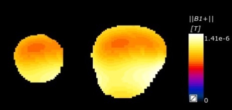
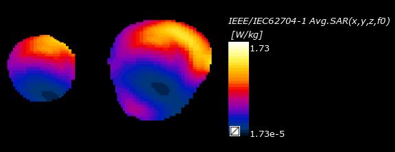
Spatial variations of B1+ and SAR10g in the central axial plane within fetus and uterus for model A and $$$\sigma_{AF}$$$ =1.25 S/m are shown in figures 2 and 3 respectively.
Discussion
Increasing the amniotic fluid conductivity results in a greater power requirement to achieve a chosen mean B1+ in the volume of interest, leading to increases in the SAR10g and coefficient of variation of B1+. This suggests that models in which the commonly adopted8,9 approach of using $$$\sigma_{CSF}$$$ as a surrogate for $$$\sigma_{AF}$$$ may lead to an overestimation of local SAR. Since $$$\sigma_{AF}$$$ falls by ~10% beyond ~ 30 weeks10 and the AF volume reaches maximum at ~32 weeks and then decreases, attenuation will reduce with time. Results from adjusting the fetal dielectric properties relative to those of adult tissues due to the increased water content (~80% cf ~55% at 28 weeks) as reported in the literature5–8,10 suggest that reliance on adult based values may underestimate fetal local SAR10g maxima by approximately 20-30%, a greater difference than that suggested previously11 . However, a more extensive database of measured age dependent fetal properties is required to support predictions based on mixture equations. The differences obtained between the detailed and homogeneous fetal models suggest a smaller change in predicted SAR10g on the order of 10%. This study also suggests a need to account for fetal movement in assessing RF safety, in accord with Murbach et al11. In the example investigated a small change of 17 mm in fetal position (5mm posterior and 13 mm to the right relative to fig 1) resulted in a change of up to 5% in SAR10g in fetal tissue.Conclusion
This investigation into the effect of uncertainty in dielectric properties on B1+ and SAR predictions for fetal MRI suggests that (a) an accurate value of $$$\sigma_{AF}$$$, rather than a surrogate will avoid overestimating power to maintain a chosen B1+ value in the fetus and fetal SAR10g, (b) fetal properties adjusted for gestational age impact on B1+ and result in significant increase in fetal SAR10g compared to adult value based simulations, (c) smaller changes in SAR10g are predicted between detailed fetal models and homogeneous ones with volume weighted mean dielectric properties, (d) fetal movement should also be taken into account.Acknowledgements
SJM acknowledges Fellowship support from the EPSRC (EP/L00531X/1)
The research was supported by the National Institute for Health Research (NIHR) Biomedical Research Centre based at Guy's and St Thomas' NHS Foundation Trust and King's College London. The views expressed are those of the authors and not necessarily those of the NHS, the NIHR or the Department of Health.
References
1. Gosselin, M.-C. et al. Development of a new generation of high-resolution anatomical models for medical device evaluation: the Virtual Population 3.0. Phys. Med. Biol. 59, 5287–5303 (2014).
2. IT’IS database for thermal and electromagnetic parameters of biological tissues, v3.0. (2015). doi:10.13099/VIP21000-03-0
3. Peyman, A., Gabriel, C., Benedickter, H. R. & Fröhlich, J. Dielectric properties of human placenta, umbilical cord and amniotic fluid. Phys. Med. Biol. 56, N93-8 (2011).
4. De Luca, F., Cametti, C., Zimatore, G., Maraviglia, B. & Pachì, A. Use of low-frequency electrical impedance measurements to determine phospholipid content in amniotic fluid. Phys. Med. Biol. 41, 1863–9 (1996).
5. Peyman, A. & Gabriel, C. Dielectric properties of rat embryo and foetus as a function of gestation. Phys. Med. Biol. 57, 2103–2116 (2012).
6. Wang, J., Fujiwara, O. & Watanabe, S. Approximation of aging effect on dielectric tissue properties for SAR assessment of mobile telephones. Electromagn. Compat. IEEE Trans. 48, 408–413 (2006).
7. Smith, S. R. & Foster, K. R. Dielectric properties of low-water-content tissues. Phys. Med. Biol. 30, 965–973 (1985).
8. Dimbylow, P. SAR in the mother and foetus for RF plane wave irradiation. Phys. Med. Biol. 52, 3791–3802 (2007).
9. Hand, J. W., Li, Y., Thomas, E. L., Rutherford, M. a & Hajnal, J. V. Prediction of specific absorption rate in mother and fetus associated with MRI examinations during pregnancy. Magn. Reson. Med. 55, 883–93 (2006).
10. Varsier, N. et al. Influence of pregnancy stage and fetus position on the whole-body and local exposure of the fetus to RF-EMF. Phys. Med. Biol. 59, 4913–4926 (2014).
11. Murbach, M. et al. Pregnant women models analyzed for RF exposure and temperature increase in 3T RF shimmed birdcages. Magn. Reson. Med. (2016).
Figures