5561
Contrast Deposition Within the Dentate Nucleus After Repetitive Administration: Comparison of Linear versus Macrocyclic Gadolinium Contrast Agents1Department of Radiology & Biomedical Imaging, University of California, San Francisco, San Francisco, CA, United States
Synopsis
Although gadolinium deposition in nephrogenic systemic fibrosis is limited to patients with abnormal renal function, more recent rat and human studies have demonstrated gadolinium deposition in specific brain structures in subjects without renal failure. We report here one of the largest studies evaluating gadolinium deposition in terms of both number of patients and number of doses, verifying the impact of the structural differences between the two most commonly used classes of agents (linear vs macrocyclic) and demonstrating a previously undescribed plateau in signal intensity ratio increase in patients who have received more than 25 administrations of gadolinium.
Purpose
To evaluate and compare differences in gadolinium retention between gadolinium contrast agent classes (linear vs macrocyclic) through the measurement of signal intensity in the dentate nucleus and globus pallidus. The effect of numerous administrations (10+) will also be evaluated.Methods
Institutional review board compliant retrospective review of imaging records was used to identify 77 patients with multiple gadolinium-enhanced MRI scans of the brain utilizing only one type of gadolinium contrast agent. 76 patients were imaged for malignancy follow-up, and one for history of nocardia abscess. The lowest estimated glomerular filtration rate at which a patient received gadolinium was 44. 45 patients received only gadopentetate dimeglumine (Magnevist, linear) while 32 patients received only gadobutrol (Gadavist, macrocyclic). Measurements of signal intensity were obtained of the dentate nucleus, pons, globus pallidus, and thalamus on unenhanced T1-weighted sequences. These measurements were used to calculate the dentate nucleus to pons (DNP) and globus pallidus to thalamic (GPT) signal intensity ratios for the patient’s initial and last available MRI studies. T-tests were used to analyze the difference in DNP and GPT ratios between the first and last exams.Results
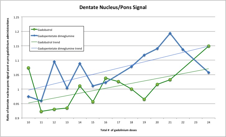
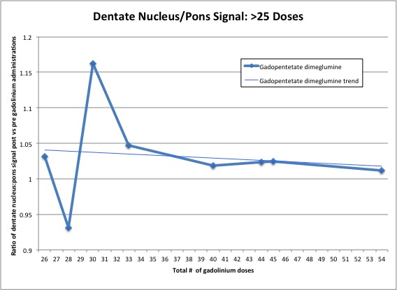
Patients receiving gadopentetate dimeglumine (linear) received an average of 19.7 contrast-enhanced MR exams, 49% were male, had a mean age of 46.5 years old at first exam, and received their last exam an average of 1163 days after the initial study. The 32 patients receiving gadobutrol (macrocyclic) had an average of 15.4 MR contrast-enhanced exams, 50% were male, had a mean age of 50.3 years at time of first exam, and received their last exam an average of 851 days after the initial study. No patients received over 24 doses of gadobutrol. However, 9 patients received 25 or more doses of gadopentetate dimeglumine. The highest number of administrations in a single patient was 54 gadopentetate dimeglumine contrast-enhanced exams. There was a significant change in mean DNP ratio with gadopentetate dimeglumine (linear) increasing from 1.06 to 1.1 (p=0.0008). Increases in DNP ratio with gadobutrol (macrocyclic) administration were not statistically significant with a mean DNP ratio before and after contrast administration of 1.06.Discussion
Increasing dentate nucleus signal after administration of gadolinium agents has been described (Kanda et al 2014, Errante et al 2014, Ramalho et al 2015, Cao et al 2016). Most studies have involved small numbers of patients or low numbers of administrations (≤6 doses). With an overall average of 17 administrations, this study is one of the largest studies verifying a detectable and statistically significant amount of gadolinium deposition with a linear contrast agent (gadopentetate dimeglumine) and confirming no significant gadolinium deposition in the dentate nucleus with a macrocyclic agent (gadobutrol).
The increase in DNP ratio seen in patients with less than 24 total administrations matches the data obtained in prior studies. However, in the group of 9 patients with more than 25 administrations of the linear contrast agent, a plateau in DNP increases was identified. No continued elevation in DNP was seen, even after 54 administrations.
In patients with fewer than 25 doses of gadolinium, with increasing number of gadolinium doses there is a trend towards an increase in DNP ratio post-gadolinium compared to pre-gadolinium. Although this was only statistically significant in the linear contrast agent, this overall trend applies to both linear and macrocyclic gadolinium agents. In our patients with more than 25 doses of a linear gadolinium agent, there is a non-statistically significant trend towards a lower increase in DNP ratio post-gadolinium compared to pre-gadolinium. The variability in DNP ratio change in patients receiving greater numbers of doses confirms the complexity of this issue. These findings suggest there are multiple other factors including specific physiologic or tissue factors that could be affecting both the deposition and/or clearance of gadolinium agents. Additional studies are needed to clarify the pathophysiology underlying gadolinium deposition as well as investigate any long-term effects.
Conclusion
Our study confirms the statistically significant increase in dentate nucleus signal following administration of linear gadolinium agents and the lack of statistically significant signal increase with macrocyclic agents, even with higher numbers of administered doses (10+). Our study also found that the number of doses was not a clear predictor of DNP increase, with the patients who received the highest number of doses not demonstrating the greatest increase in DNP ratio. These findings highlight the complexity of gadolinium deposition and demonstrate the need to further larger, longitudinal studies monitoring not only contrast deposition but also clinical implications.Acknowledgements
No acknowledgement found.References
Kanda T, Ishii K, Kawaguchi H, Kitajima K, Takenaka D. High signal intensity in the dentate nucleus and globus pallidus on unenhanced T1-weighted MR images: relationship with increasing cumulative dose of a gadolinium-based contrast material. Radiology. 2014;270(3):834-41.
Errante Y, Cirimele V, Mallio CA, Di lazzaro V, Zobel BB, Quattrocchi CC. Progressive increase of T1 signal intensity of the dentate nucleus on unenhanced magnetic resonance images is associated with cumulative doses of intravenously administered gadodiamide in patients with normal renal function, suggesting dechelation. Invest Radiol. 2014;49(10):685-90.
Ramalho J, Castillo M, Alobaidy M, et al. High Signal Intensity in Globus Pallidus and Dentate Nucleus on Unenhanced T1-weighted MR Images: Evaluation of Two Linear Gadolinium-based Contrast Agents. Radiology. 2015;276(3):836-44.
Cao Y, Huang DQ, Shih G, Prince MR. Signal Change in the Dentate Nucleus on T1-Weighted MR Images After Multiple Administrations of Gadopentetate Dimeglumine Versus Gadobutrol. AJR Am J Roentgenol. 2016;206(2):414-9.
Figures
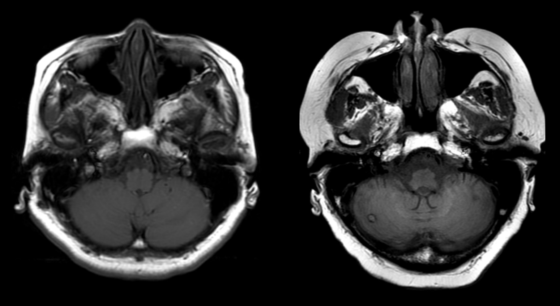
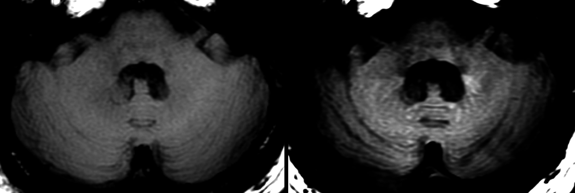
Axial un-enhanced T1 images through the posterior fossa demonstrating the increase in signal intensity believed secondary to gadolinium accumulation.