5556
Quantitative Image Analysis of Real-Time Golden Angle Radial iSSFP for Interventional MRI1Radiological Sciences, University of California Los Angeles, Los Angeles, CA, United States, 2Biomedical Physics, University of California Los Angeles, Los Angeles, CA, United States, 3Bioengineering, University of California Los Angeles, Los Angeles, CA, United States
Synopsis
Real-time visualization is crucial to the success of MRI-guided minimally invasive cancer interventions. We have developed golden-angle (GA) ordered radial integrated-SSFP (iSSFP), which can suppress banding artifacts associated with bSSFP while maintaining similar T2/T1 contrast. In this work, we further analyze the tissue contrast as well as passive visualization of interventional needles using GA radial iSSFP. In volunteer scans, we verify that GA radial iSSFP achieves T2/T1 tissue contrast similar to bSSFP while suppressing banding artifacts. With phantom scans we show that iSSFP reduced the size of the needle-induced signal void, versus that seen on bSSFP. These advantages of GA Radial iSSFP show its potential for improving real-time MRI-guided interventions.
Introduction
Real-time visualization is crucial to the success of MRI-guided minimally invasive cancer interventions, especially in the abdomen. The real-time MRI sequence must be ultrafast, while maintaining high soft-tissue contrast, particularly T2 contrast. RF-spoiled gradient echo (GRE) is able to achieve real-time imaging speeds, but the signal-to-noise ratio (SNR) and tissue contrast is diminished. Balanced-SSFP (bSSFP) has better T2/T1 contrast and SNR but suffers from banding artifacts1. We have developed golden-angle (GA) ordered radial integrated-SSFP (iSSFP)2, which is a special case of steady-state MRI that can suppress banding artifacts associated with bSSFP while maintaining similar T2/T1 contrast. In this work, we further analyze the tissue contrast as well as passive visualization of interventional needles using GA radial iSSFP.Methods
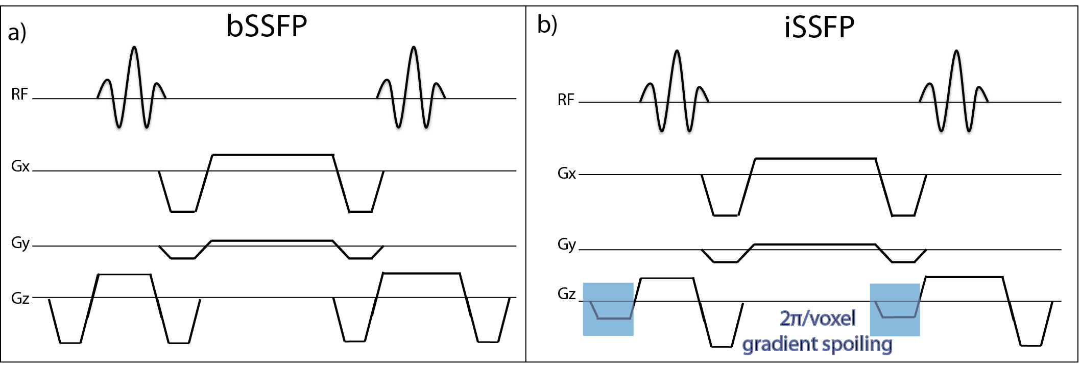
Real-time Golden-Angle Radial iSSFP Sequence: To create a GA radial iSSFP sequence, the slice select gradient of the bSSFP sequence was modified to achieve 2π/voxel gradient spoiling (Fig 1). The radial readouts were continuously acquired with GA ordering3. The GA radial iSSFP data was reconstructed using a combined sliding-window SPIRiT4 algorithm.
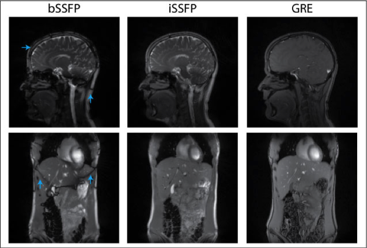
Tissue Contrast Evaluation Experiments: Volunteer experiments were performed on a 3.0T MRI scanner (Prisma, Siemens) with 300-mm field-of-view, spatial resolution of 1.5x1.5x5mm, and 4095 radial readouts. Volunteers’ brain and abdomen (breath held) were scanned using the GA radial bSSFP, iSSFP, and GRE sequences (bSSFP and iSSFP: TE/TR=2.01/4.79 ms, FA=70o; GRE: TE/TR=1.63/3.14 ms, FA=20o).
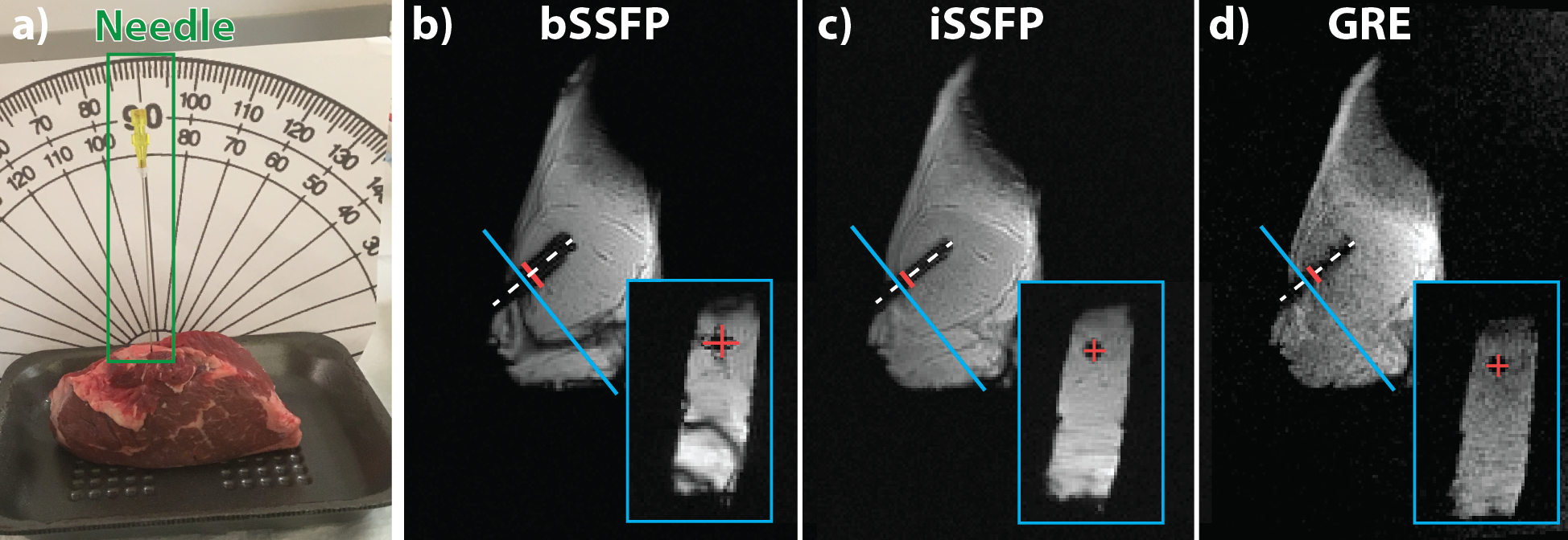
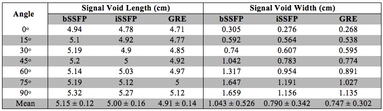
Needle Visualization Evaluation Experiment: A slab of beef steak was imaged with a 20 Gauge MRI-compatible needle (Cook Medical MReye Disposable Chiba Biopsy Needle) inserted 4.6cm at a range of angles (0 to 90o, in steps of 15o) with respect to the B0 field (Fig 2a). The setup was scanned using the GA radial bSSFP, iSSFP, and GRE sequences (bSSFP and iSSFP: TE/TR=2.14/6.02ms, FA=50o; GRE: TE/TR=1.74/3.93ms, FA=50o), with 258-mm field-of-view, spatial resolution of 1.3x1.3x3.5mm, and 4095 radial readouts.
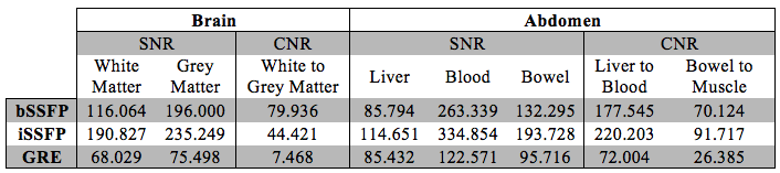
Image Reconstruction and Analysis: In order to facilitate inter-sequence SNR and CNR comparison in corresponding ROIs, we maintained consistent imaging position by use of breath-held and static scans. For the brain images, the SNR of the white matter, grey matter and cerebrospinal fluid (CSF), as well as the white to grey matter CNR was computed. For the abdominal images, the SNR of the liver, blood, and bowel, and the CNR of the liver to blood, and bowel to muscle was computed. For the needle experiments, the length and the width of the signal void from the needle was measured along each plane for radial bSSFP, iSSFP, and GRE images (Fig 2).
Results
Tissue Contrast: The SNR and CNR results are listed in Table 1. As expected iSSFP has similar SNR and CNR results as bSSFP and also eliminates banding artifacts (Fig 3) in both the brain and abdomen. Compared to GRE, iSSFP had over three fold higher CNR in all cases.
Needle Visualization: The images for all insertion angles were viewed for banding artifacts. It can be seen that bSSFP has banding artifacts, both at the edge of the meat and at the needle tip (Fig 2). The measured signal void length was averaged for all the planes and insertion angles (Table 2), and was compared to the known insertion length of 4.6 cm. The measured signal void width was also was averaged for all the planes and insertion angles, and compared to the known needle width of 0.9 mm. On average iSSFP had reduced signal void lengths and widths compared to bSSFP, and was comparable to GRE.
Discussion and Conclusion
Our GA radial iSSFP sequence combines the tissue contrast benefits of the bSSFP sequence without the banding artifacts, which can potentially improve real-time MRI-guided interventions. . In volunteer scans at 3.0T, it is verified that GA radial iSSFP achieves sufficient T2/T1 tissue contrast, similar to bSSFP, while suppressing banding artifacts. In addition, iSSFP reduced the size of the needle-induced signal void versus that seen on bSSFP, which will improve the physician visualization of the location of the needle and the surrounding tissue. These advantages of GA radial iSSFP show its potential for improving real-time MRI-guided interventions.Acknowledgements
This project was supported in part by an NSF Graduate Research FellowshipReferences
1. Hargreaves BA. Rapid gradient-echo imaging. J. Magn. Reson. Imaging [Internet] 2012;36:1300–13. doi: 10.1002/jmri.23742.
2. Mikaiel S, Martin T, Sung K, Wu H. Real-Time Golden Angle Radial iSSFP for Interventional MRI. In: Proceedings 24th Scientific Meeting, International Society for Magnetic Resonance in Medicine. ; 2016. p. 3579.
3. Winkelmann S, Schaeffter T, Koehler T, Eggers H, Doessel O. An optimal radial profile order based on the Golden Ratio for time-resolved MRI. IEEE Trans. Med. Imaging [Internet] 2007;26:68–76. doi: 10.1109/TMI.2006.885337.
4. Lustig M, Pauly JM. SPIRiT: Iterative self-consistent parallel imaging reconstruction from arbitrary k-space. Magn. Reson. Med. [Internet] 2010;64:457–71. doi: 10.1002/mrm.22428.
Figures