5545
MR-Guided Mixed-Reality For Surgical Planning: Set-Up and Perceptual Accuracy1Department of Radiological Sciences, Stanford University, Palo Alto, CA, United States, 2Department of Surgery, Stanford University, Palo Alto, CA, United States
Synopsis
Microsoft HoloLens provides the ability to visualize 3D holograms of preoperative MRI in addition to the physical environment. In this work we have developed a HoloLens application that aligns these preoperative holograms to the patient. The accuracy of perceiving these holograms was evaluated by presenting different shapes of holograms in random locations and comparing their positions to ground truth. The current set-up enables visualization and perception of holograms with a margin tolerance of < 6 mm.
Introduction
Three-dimensional high resolution MRI enables visualization of patient anatomy with sufficient tissue contrast. However, conventional preoperative MRI and even 3D rendered MRI are presented to the surgeon on a stand-alone monitor that is separate from the patient, and requires the surgeon to mentally translate findings to the patient when planning surgery. The ability to visualize 3D MRI overlaid onto the patient would enable surgeons to better visualize the anatomy and potentially improve surgical planning. In this work, we have: (i) developed a mixed-reality HoloLens application to visualize the ‘hologram’ of MRI registered to the patient; and (ii) evaluated the perceptual accuracy of the ‘holograms’ rendered by the HoloLens.Methods
Microsoft HoloLens1 enables mixed-reality by projecting ‘holograms’ into the visual field. This will enable a surgeon to view the patient as well as ‘holograms’ of pre-operative or diagnostic MRI projected onto the patient. The ‘holograms’ can be registered and locked to the patient using fiducials that are both MR and optically visible, enabling the surgeon to move around the patient for 3D visualization of the MRI from different angles.
HoloLens Application Functionalities: An initial application was developed in HoloLens and tested on a torso phantom. Three-dimensional, spoiled gradient-echo MR images of a torso phantom were acquired with 5 gadolinium filled optical fiducials as shown in Figure 2. The center positions of the markers in the 3D MRI were measured using Osirix2. A contrast-enhanced 3D breast MRI dataset3 was warped to fit the torso phantom4. Skin, chest and a breast tumor were segmented semi-automatically5 and the corresponding surface meshes were created. The ‘holograms’ of the MRI slices can be viewed along with the meshes in different viewing planes (sagittal, coronal, transverse and cut-away) using voice commands (Figure 2). Different slices can be scrolled over using the gesture controls of HoloLens. OpenCV6,7 was integrated in HoloLens and computer vision algorithms were used for automatic detection of the fiducials and registration of the holograms to the phantom. The ‘holograms’ can also be manually registered by either: (1) translating and/or rotating about a plane of the one or more fiducials; or (2) using the user’s head position to move the holograms.
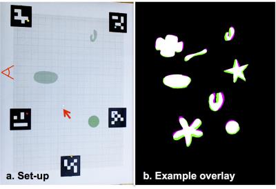
Perceptual Accuracy: A graph paper sheet with five fiducials at the corners (Fig. 3a) was used to evaluate the 2D perceptual accuracy of holograms by 6 subjects. The HoloLens was initially set up to the measured inter-pupillary distance (IPD) of the subject. Later, each subject calibrated the holograms of the fiducials to the automatically detected fiducials in the graph paper. This user-specific calibration was essential to know the position of the HoloLens with respect to the eyes of the subject. The subjects were then presented with 1mm spherical holograms and were asked to draw each location in the graph paper. The error between the marked positions of 20 such spheres located in random positions, and the ground truth was measured. The subjects were also presented with 8 holographic shapes with longest dimension varying from 2 to 4.5 cm and their perceptual accuracy (Fig. 3b) was also analyzed.
Results and Discussion
The HoloLens application is able to register and lock the MRI to the rigid torso phantom using the MR-visible optical fiducials as shown in Figure 2. In the future, 3D perceptual accuracy of palpable tumors will be evaluated with contrast-enhanced supine MRI. Deformable registration of acquired supine MRI to the patient in surgical position will also be considered.
The errors between the marked spheres or shapes, and the ground truth are tabulated in Figure 3. The margin tolerance, the number of additional dilated pixels required on the drawn shapes to completely cover the ground truth shapes, ranged between 0.7 to 5.7 mm. The range of errors along the depth (horizontal) direction, was larger compared to the vertical direction. This could be because the perceptual accuracy of the holograms, in addition to the accurate alignment of the holograms to the object, depends on various factors including IPD, position of the HoloLens in the head. These additional parameters were adjusted during user-specific calibration. Further improvements in calibration are being considered to further improve perceptual accuracy.
Conclusion
A preliminary application in HoloLens has been created to visualize the holograms of three-dimensional MR slices aligned to a torso phantom. The application enables visualization and perception of holograms with a margin tolerance of < 6 mm, which should be improved further with more advanced user-specific calibration methods.Acknowledgements
Stanford Cancer Imaging Training Program T32 CA 009695
Microsoft Corporation
California Breast Cancer Research Program IDEA award 22IB-0006
References
1. www.microsoft.com/microsoft-hololens/
2. http://www.osirix-viewer.com/
3. https://wiki.cancerimagingarchive.net/display/Public/QIN+Breast+DCE-MRI
4. www.slicer.org
5. Yushkevich PA et al., Neuroimage 2006;31(3):1116-28 (www.itksnap.org/)
6. http://opencv.org/
7. Garrido-Jurado et al., Pattern Recogn. 2014; 47(6):2280-2292
Figures


