5544
Inline Adaptive Spiral Off-Resonance Correction for MRI-guided interventions1Biochemistry and Biophysics Center, Division of Intramural Research, National Heart, Lung, and Blood Institute, National Institutes of Health, Bethesda, MD, United States
Synopsis
Spiral imaging is appealing for MRI-guided interventions due to the need for high frame rate dynamic imaging and low RF power sequences to reduce RF-induced heating in metallic guidewires. Unfortunately, spiral images are susceptible to image distortions due to off-resonance which must be corrected. In this work, we implement a real-time interactive spiral sequence and a fast reconstruction in the Gadgetron that performs inline off-resonance correction that adapts to slice position and orientation changes. We show the effectiveness of our correction in both phantom and in-vivo volunteer images with reconstruction times that are approaching real-time.
Introduction
MRI-guided interventions require imaging sequences that are fast, interactive and RF-efficient. Spiral imaging is particularly appealing for high frame rate dynamic imaging in the interventional MRI environment and has been demonstrated to reduce RF-induced heating in metallic guidewires.1 Unfortunately, spiral images are susceptible to image distortions due to off-resonance which must be corrected inline in real-time in the context of interventional MRI to produce a live stream of high quality images for procedural guidance. Previously, we have shown real-time correction of gradient distortion in spiral images2, and here we address the real time off-resonance correction (deblurring) of spiral images. In this work, we implement a real-time interactive spiral sequence and reconstruction that performs inline off-resonance correction that adapts to slice position and orientation changes.
Methods
All images were acquired using a 1.5T scanner (Aera, Siemens, Erlangen, Germany). We adapted our real-time spiral sequence to begin with two short single shot spirals acquired with a 1 ms TE offset from which we compute an off-resonance map (allowing for off-resonant frequencies between +/- 500 Hz to be resolved without wrapping). Slice geometry is updated frequently during an MRI-guided procedure and, therefore, an off-resonance map is re-acquired for every change of slice orientation or position. A low pass Gaussian filter is applied to the single shot spiral acquisitions to suppress high spatial frequency off-resonance components that are subject to breathing and cardiac motion. Following acquisition of the off-resonance map, the real-time spiral sequence continues to acquire a stream of multi-shot spiral images.The spiral reconstruction and off-resonance correction ran on a remote computer with a GPU using the Gadgetron open source image reconstruction framework3, written in C++. While scanning, the de-blurring “Gadget” continuously received data one spiral readout at a time. If the gadget detected a single shot acquisition, a new off-resonance map was computed and stored in memory. For each real-time multishot spiral image, a series of base images were computed in a range of equally spaced demodulation frequencies. Multi-frequency interpolation (MFI) was used to compute a fast conjugate phase reconstruction image based on the off-resonance map4. MFI used predetermined coefficients, stored as a look-up table, to weight and sum the series of demodulated base images. The Gadget pipeline is illustrated in Figure 1.
The inline adaptive spiral off-resonance correction was tested in a phantom and in a healthy volunteer with the following parameters: TE/TR = 0.86/5.16 ms (16-shot spiral) or 0.86/8.6 ms (8-shot spiral), flip angle = 10°, FOV = 300x300 mm, matrix size = 128x128.
Results
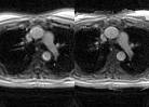
Phantom experiments showed that our inline off-resonance strategy adequately compensates for slow spatially varying off-resonance induced by a linear B0 gradient (Fig 2). The adaptation of the off-resonance map to changing slice orientation is animated in Fig 3 where a linear B0 gradient is present in 2 axes. The adaptive sequence that recalculates the off-resonance map for each slice geometry change maintains off-resonance correction for arbitrary image orientation. The real-time off-resonance correction was tested in a normal volunteer (Fig. 4&5), producing clear improvement of spiral image blurring at vessel edges and tissue boundaries.
The number of base images needed for MFI depends on the length of each readout (interleave). An 8-shot sequence requires 13 MFI base images and reconstructs in an average time of 445 ms per image. Similarly, a 16-shot sequence requires 7 base images and reconstructs in 300 ms per image. The map reconstruction takes an additional 410 ms.
Discussion
Here we have demonstrated inline, adaptive reconstruction of off-resonance corrected spiral images. The reconstruction times are fast, but not yet real-time as needed for interventional MRI. Future work will be aimed at using multiple GPUs and compute the MFI images in parallel. In an interventional setting, multiple slices are often acquired in simultaneously. These slices can be reconstructed on separate nodes to further improve reconstruction speed. The Gadgetron framework is vendor-agnostic and allows leveraging of high power computational resources, such as GPUs and distributed cloud computing, for inline reconstruction.
The remaining blurring in the in-vivo images is likely due to changes in the off-resonance field with motion. Further improvement could be gained by adding an automatic off-resonance map generation stage5,6, although this would add computational demand. This correction method will also be tested in the presence of a metallic guidewire, which would add a significant additional susceptibility related off-resonance.
Conclusion
Real-time off-resonance correction is essential to improve the quality of the spiral images used for MRI-guided interventions. We have shown here an adaptive real-time sequence and fast inline off-resonance corrected reconstruction.
Acknowledgements
No acknowledgement found.References
1. Campbell-Washburn AE et al. Positive contrast spiral imaging for visualization of commercial nitinol guidewires with reduced heating. JCMR. 2015;17:114.2. Campbell-Washburn AE et al. Real-time distortion correction of spiral and echo planar images using the gradient system impulse response function. Magn Reson Med. 2016 Jun;75(6):2278-85.
3. Hansen MS, Sørensen TS. Gadgetron: an open source framework for medical image reconstruction. Magn Reson Med 2013;69:1768–177.
4. Nayak KS, Nishimura DG. Automatic field map generation and off-resonance correction for PR imaging. Magn Reson Med 2000; 43:151–154.
5. Man LC, Pauly JM, Macovski A. Multifrequency interpolation for fast off-resonance correction. Magn Reson Med 1997; 37:785–792.
6. Chen W, Meyer CH. Semiautomatic off-resonance correction in spiral imaging. Magn Reson Med 2008;59:1212–1219.
Figures
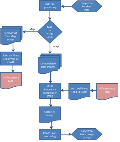
Figure 1: Gadgetron reconstruction pipeline used for inline reconstruction of deblurred spiral images. The header identifies data as belonging to the off-resonance map acquisition or to the multi-shot interleaved image. The off-resonance map is highlighted in a different color to show that it is saved and recalled from memory – instead of being passed down the pipeline linearly. This is important because the same map is used repeatedly until the imaging slice/orientation changes and a new map is acquired.
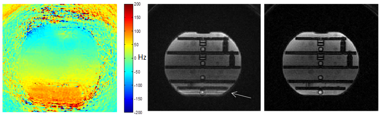
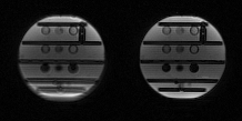
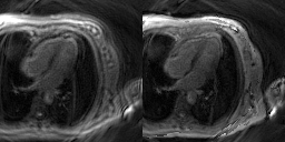
Figure 4: 8-shot spiral images of a 4 chamber view with the same parameters as shown Fig 2. (Left) Original reconstruction. (Right) Off-resonance corrected.