5526
Prospective frequency correction using outer volume suppression-localized navigator for MR Spectroscopic Imaging1Hoglund Brain Imaging Center, University of Kansas Medical Center, Kansas City, KS, United States, 2Department of Neurology, University of Kansas Medical Center, Kansas City, KS, United States, 3Department of Molecular & Integrative Physiology, University of Kansas Medical Center, Kansas City, KS, United States
Synopsis
Data acquisitions for magnetic resonance spectroscopic imaging (MRSI) require a long scan time to increase SNR and for spatial encoding. During the prolonged scan time, maintaining a constant static magnetic field (B0) is important for a robust MRSI measurement. However, frequency drifts occur over time even in advanced MR systems and become larger when high shim currents or rapidly switched gradients are applied. The frequency drift causes broad and distorted spectral lineshapes, reduced SNR, and quantification errors. These effects can be mitigated retrospectively and prospectively. However, in MRSI measurements, these effects can only be mitigated using the prospective frequency correction, because each spectrum is phase-encoded. The prospective frequency correction is typically achieved by incorporating a PRESS-based interleaved reference scan (PRESS-IRS) as a navigator, termed as PRESS-IRS navigator. A small excitation flip angle (10-20°) is used for the PRESS-IRS navigator to reduce the saturation-induced SNR loss on metabolite signals. Nonetheless, the SNR loss remains unavoidable and becomes notable when the imperfect refocusing pulses or a short repetition time (TR) are used in MRSI. In this study, a new prospective frequency correction method is introduced. The new method utilizes the outer volume suppression-localized navigator, termed OVS-localized navigator, resulting in no perturbations of metabolite signals and thus no saturation-induced SNR losses. Meanwhile, a precise measurement of the frequency drift and the effective correction is achieved. The presented method was demonstrated in two-dimensional (2-D) MRSI measurements under the large frequency drift induced by a fMRI experiment.
PURPOSE
Data acquisitions for magnetic resonance spectroscopic imaging (MRSI) are sensitive to frequency drifts during the relatively long scan time to increase SNR for low concentrations of metabolites and for spatial encoding. Significant frequency drifts occur when high shim currents or rapidly switched gradients are applied even in advanced MR systems. 1 The frequency drifts cause broad and distorted spectral lineshapes, reduced SNR, quantification errors, and errors in spatial encoding. 2-12 Although these effects can be mitigated retrospectively, prospective frequency correction methods are more appropriate for MRSI because MRSI lacks identifiable reference MRS signals due to phase-encoding. 2,6,8 The prospective frequency correction is typically achieved by incorporating a PRESS-based interleaved reference scan (PRESS-IRS) as a navigator. 4,6,8 Small flip angle (10-20°) excitation pulses 4,6,8 are generally used for the PRESS-IRS navigator to reduce the saturation-induced SNR loss on metabolite signals. Nonetheless, the SNR loss remains unavoidable and becomes notable with the imperfect refocusing pulses or a short repetition time (TR). In this study, we introduce a new prospective frequency correction method with a navigator that uses outer volume suppression (OVS)-localization and selective water excitation, which overcomes major shortcomings of previous methods.METHODS
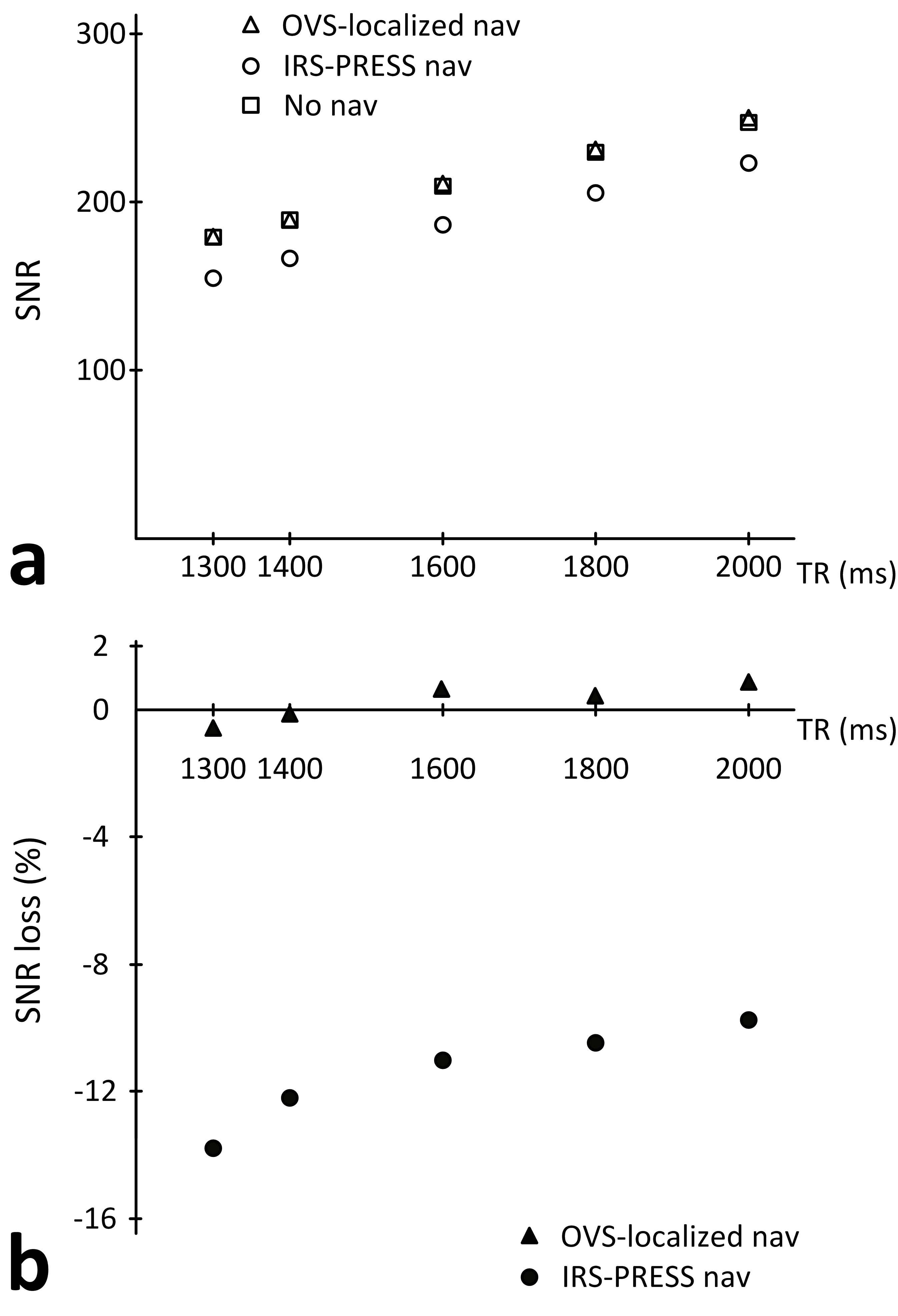
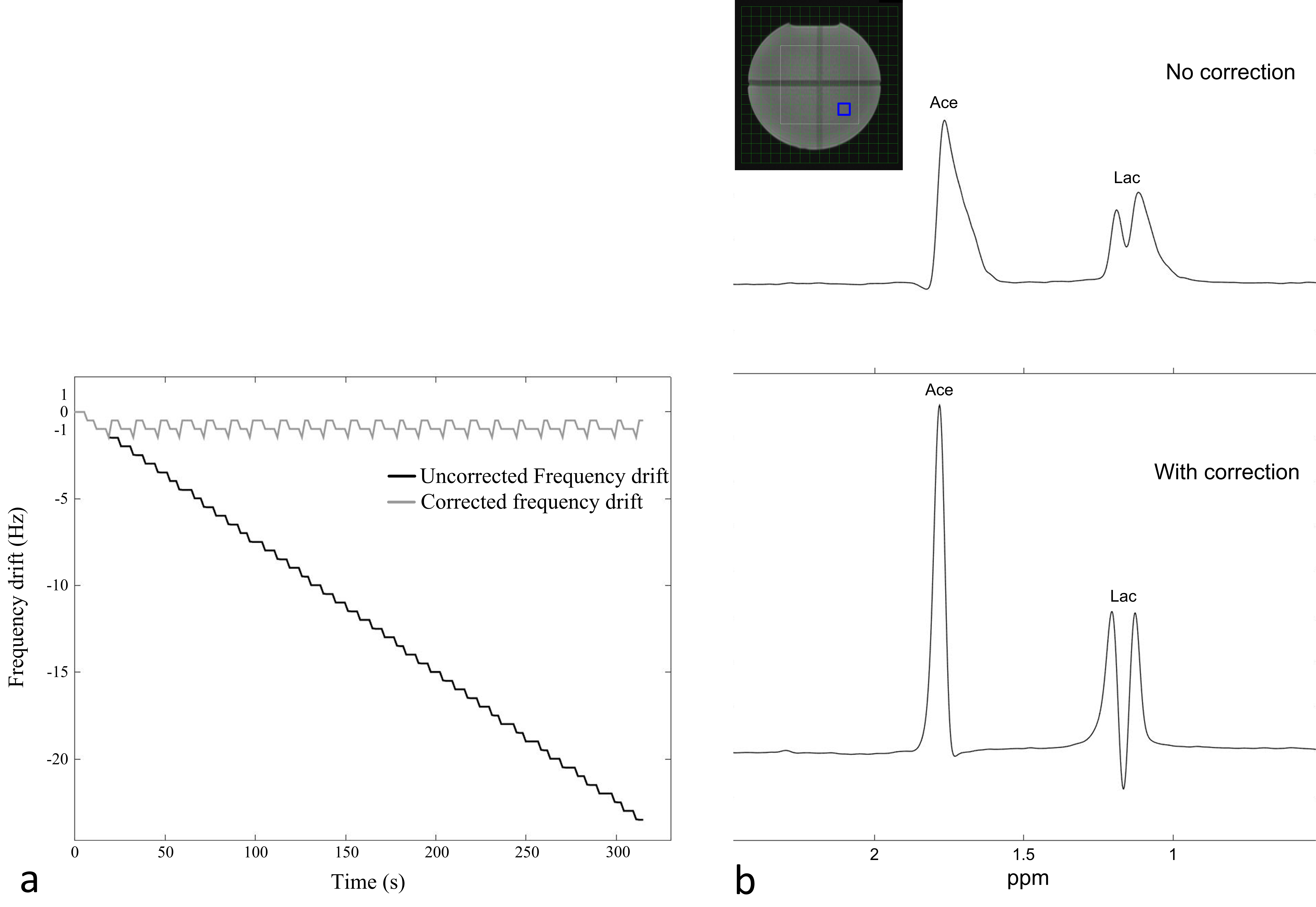
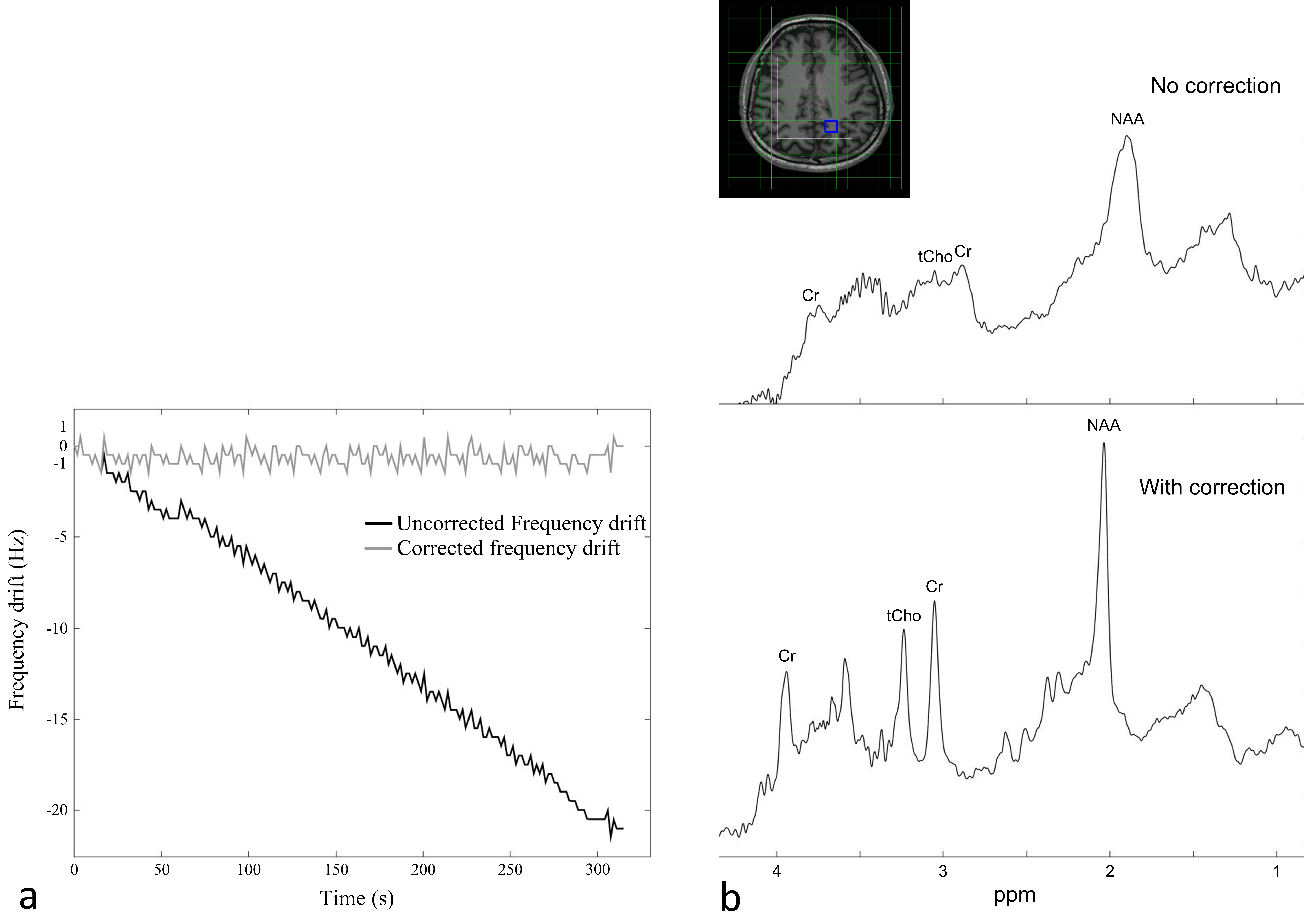
The proposed OVS-localized navigator method incorporates a navigator acquisition with the OVS localization 13,14 and selective water excitation into the MRSI data acquisition (Fig. 1). The OVS localization was adjusted to match the volume localization in PRESS MRSI. The localized water signal was acquired and sent to the real-time reconstruction system, where the peak position of the water signal was used to determine the frequency drift. The measured frequency drift was then used to prospectively update the frequencies of the RF pulses and the receiver for data acquisition at the next acquisition. The OVS-localized navigator method was developed and integrated into a semi-LASER based single-voxel MRS and 2D MRSI sequences 15 on a 3 T scanner (Skyra, Siemens, Erlangen, Germany). To examine the effectiveness of our proposed method, the PRESS-IRS navigator method with an excitation flip angle of 15° was also implemented in the single-voxel MRS sequence. At the first experiment, the OVS-localized navigator method was compared with the PRESS-IRS navigator method with and without a navigator acquisition for the SNR. The SNR comparisons were performed using the single-voxel MRS measurements on a phantom containing solutions of acetate and lactate. The experiment was repeated five times. The SNR differences were assessed using the two-sample t-test with a significance level p = 0.05. At the second experiment, the OVS-localized navigator method was demonstrated in 2-D MRSI measurements following a 50-min fMRI experiment to create realistic gradient heating-induced frequency drifts. The experiment was performed on a healthy volunteer and a phantom containing solutions of acetate and lactate. Parameters for the 2-D MRSI sequence were: FOV = 20×20 cm2, thickness = 2 cm, VOI = 10×10 cm2 in a phantom and 9×9 cm2 in humans, 16×16 phase encodings, TE/TR = 35/1700 ms, and bandwidth = 2000 Hz.RESULTS
The SNR using the OVS-localized navigator method showed no statistical differences compared to the SNR without the navigator (p = 0.55-0.95) (Fig. 2a). The SNR differences were less than 1 % across TRs (Fig. 2b). However, the SNR using the IRS-PRESS navigator method was significantly lower than the SNR without the navigator (p = 1.5 × 10⁻⁴-3.5 ×10⁻⁶). The saturation-induced SNR losses were 9.7-13.7% and became more notable at a shorter TR. In phantom and human MRSI measurements (Figs. 3 and 4), the fMRI experiment-induced frequency drifts were similar with the rate of approximately -4.5 and -4.1 Hz/min and the maximums of -23.5 and -21.5 Hz, respectively. The measured frequency drifts in the human brain showed a larger fluctuation than in the phantom, which may be due to physiological motions and increased uncertainties in determining the peak position in the human. The frequency drifts resulted in broad and distorted lineshapes of MRSI spectra (Figs. 3 and 4). By using the OVS-localized navigator method, the maximal frequency drift was reduced to -1.5 Hz, and the effects of frequency drifts were notably reduced. Thus, the spectra acquired with the frequency correction showed enhanced linewidths and better-defined metabolite signals, including sharper acetate and lactate peaks in the phantom, and NAA, creatine, glutamate/glutamine and choline peaks in the human.Acknowledgements
No acknowledgement found.References
No reference found.Figures