5507
T2 relaxation times of metabolites measured with LASER and PRESS at 3 TDinesh K Deelchand1, Edward J Auerbach1, and Małgorzata Marjańska1
1CMRR, University of Minnesota, Minneapolis, MN, United States
Synopsis
The goal of this study was to compare the apparent transverse relaxation time (T2) constants of metabolites obtained using LASER and PRESS sequences in the human brain at 3 T. A 25% higher apparent T2s of total N-acetyl aspartate, total creatine and total choline were measured with LASER sequence as compared to PRESS while comparable apparent T2s were measured for strongly coupled metabolites, e.g., glutamate and myo-inositol, with both sequences.
Purpose
Accurate quantification of proton spectra acquired at relatively long echo-time (TE) requires the knowledge of T2 relaxation time constants in order to correct for signal losses. Several studies, both in humans1 and animals2, have shown that the apparent T2 rates of singlet resonances such as tCr, tNAA and tCho are increased under the effect of a Carr-Purcell (CP) pulse train. In addition, we previously showed that the increase in the apparent T2 for strongly J-coupled metabolites (glutamate, glutamine, myo-inositol and taurine) is much larger (at least 2 times) compared to singlets under T2p regime2. However, so far there have not been any studies comparing the T2 values of metabolites between LASER and PRESS, sequences commonly used in the research settings. Based on findings from previous studies, we postulate that the apparent T2 of metabolites measured with LASER will be higher for singlets and J-coupled metabolites compared to PRESS since LASER consists of 3 pairs of refocusing pulses which act as a CP pulse train. The aim of this study was to measure the T2 of metabolites using LASER and PRESS in the human brain at 3 T.Methods
5 healthy subjects (21 ± 1 years) were scanned on a Siemens 3 T scanner after giving informed consent approved by the IRB. Body coil was used for excitation while the 32-channel receive-only head-coil was used for signal reception. A VOI of 25×25×25 mm3 was positioned in the prefrontal cortex using T1-weighted MPRAGE images. B0 shimming was achieved using system 3D-GRE shim (Brain shim mode). Localized spectra were acquired using both PRESS and LASER sequences. VAPOR scheme interleaved with OVS pulses was used in both sequences. Data were acquired at six different TEs (35, 140, 230, 290, 330, 400 ms in LASER and 23, 60, 140, 210, 270 and 400 ms in PRESS) to measure the T2 relaxation of metabolites. Water reference scans were also acquired for eddy-current correction. All spectra were processed in Matlab3: eddy current effect was first corrected followed by single-shot frequency and phase corrections. Spectra were analyzed with LCModel using simulated basis sets and measured macromolecule spectra. T2 values were obtained by fitting the amplitudes obtained from LCModel with an exponential fit.Results and Discussion
Proton spectra acquired from one subject at different TEs with PRESS and LASER sequences are shown in Figure 1. There is a noticeable difference in the peak heights of tNAA, tCr and tCho, mostly visible at long echo times suggesting that the apparent T2s of these metabolites are different between both sequences. Indeed, the T2 relaxation times of these non-coupled metabolites were found to be statistically longer with LASER compared to PRESS (Table 1). A previous study reported an increase of about ~75% for tNAA and tCr with CP-LASER compared to PRESS at 4 T1. However in the current study, the increase was merely ~25% on average with LASER, suggesting that the apparent T2 will be further lengthened when moving from LASER to CP-LASER as demonstrated in the rat brain2. The increase in apparent T2 under LASER could be explained by the fact that the CP train suppresses the diffusion component efficiently, since these moieties do not have exchangeable protons. The T2 values for Glu and Ins were found to be comparable between LASER and PRESS (Table 1). These values agree with previously published values at 3 T using PRESS3-5 where T2 of these metabolites ranged from 180 to 200 ms. Contrary to our hypothesis, no lengthening of the apparent T2 for strongly coupled spins were observed with LASER in this study. One possible explanation is that the duration of the CP pulse train in LASER is too short such that there is no refocusing of cross-correlation effects between different dipole–dipole interactions compared to when using CP-LASER and T2p-LASER sequences2.Conclusion
In summary, this study shows that the apparent T2 relaxation times of singlets are lengthened by ~25% under LASER compared to PRESS while similar apparent T2 values were observed for strongly J-coupled metabolites with both sequences. In conclusion, the LASER sequence seems to be more efficient in suppressing the diffusion component of non-coupled singlets (having non-exchangeable protons) compared to coupled metabolites.Acknowledgements
Supported by NIH grants: R21AG045606, P41 EB015894, P30 NS076408References
1. Michaeli et al. Magn Reson Med 2002
2. Deelchand et al. Magn Reson Med 2014
3. Deelchand, https://www.cmrr.umn.edu/downloads/mrspa/
4. Ganji el al. NMR Biomed 2011
5. Choi et al. Magn Reson Med 2006
6. Schubert et al. NeuroImage 2004
Figures
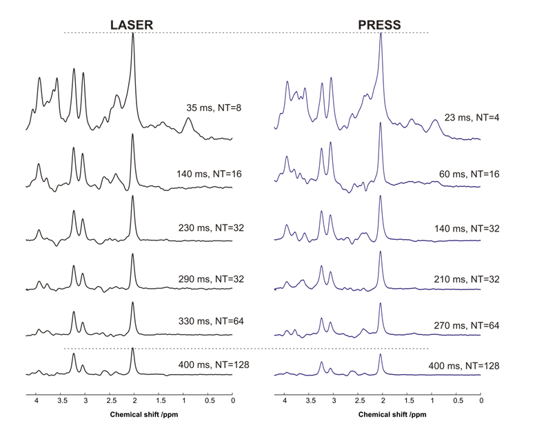
Figure 1: Spectra (TR
= 3 s with different averages, NT) acquired at several echo-times with
LASER and PRESS from the human prefrontal lobe at 3 T. For display purposes,
the spectra were normalized such that at the shortest TE, the height of NAA peak was identical. The dotted
line at 400 ms shows that the apparent T2
of NAA and other singlets are longer with LASER compared to PRESS.
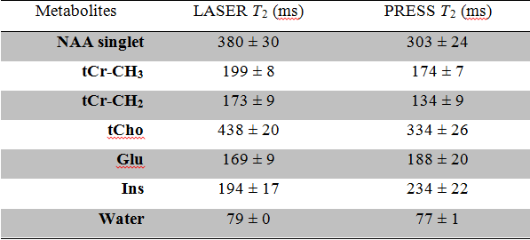
Table 1: Measured T2
values from 5 subjects using LASER and PRESS at 3 T.