5505
Overdiscrete Reconstruction for Signal Enhancement in Single Voxel Spectroscopy1Technische Universität München, Munich, Germany, 2GE Global Research, Munich, Germany, 3GE Healthcare, Potsdam, Germany
Synopsis
This work proposes an overdiscrete reconstruction for Single Voxel Spectroscopy (SVS). It is demonstrated that in single voxel acquisitions benefit from the SNR and linewidth improvement obtained by correcting for of B0 inhomogeneities and the optimization of the Spatial Response Function (SRF), as compared to regular signal averaging. This method, enables SV acquisitions in challenging brain areas, i.e. where B0 shimming is sub-optimal, and corrects for spectral artifacts such as peak aliasing.
Purpose
In this work, we propose to apply an overdiscrete reconstruction to Single Voxel Spectroscopy (SVS) to improve signal quality, reduce spectral artifacts, and increase the detection accuracy of brain metabolites and help in the detection of globally occurring metabolic changes.Introduction
SVS is a powerful tool to investigate brain function and abnormalities. A main limitation of SVS is low signal-to-noise ratio (SNR), hence requiring large voxel sizes and extensive signal averaging. However, when the voxel is large, variations of B0 field and coil sensitivity profiles cause intra-voxel dephasing and sub-optimal signal combination of multi-channel receive coils. Moreover, in many regions of the brain, susceptibility artifacts can cause peak aliasing and destroy the spectral information. The use of phase encoding at larger than the SVS resolution has demonstrated a significant suppression of these artifacts1. Recently, a reconstruction technique was introduced for MR Spectroscopic Imaging (MRSI), where stating the reconstruction problem at a resolution higher than nominal can lead to SNR improvements due to prior knowledge stemming from coil sensitivities and B0 magnetic field inhomogeneities2,3,4.Methods
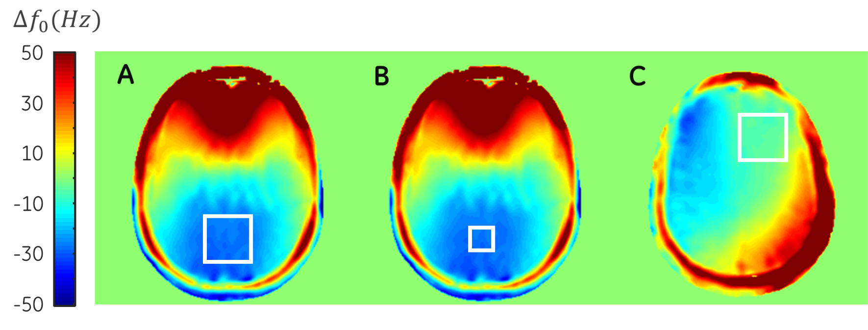
The overdiscrete reconstruction problem consist in finding the reconstruction matrix F and apply it to each temporal point of the acquired signal, where $$F^{'} = TE^{H} (EE^{H} + \alpha \Psi)^{+},$$ where $$$T$$$ represents the target SRF matrix and $$$E$$$ the encoding matrix, both expressed at $$$\zeta^{2}$$$-fold of the nominal resolution, $$$\Psi$$$ represents the noise covariance matrix, $$$\alpha$$$ is the regularization parameter to control the noise optimization and $$$^{+}$$$ denotes the pseudo-inverse. Correction for signal dephasing due to B0 inhomogeneities is applied in an intermediate step by applying the phase correction factor $$$e^{-i2 \pi\cdot t \cdot \triangle f_{0}(\mathbf r)}$$$, where $$$\triangle f_{0}(\mathbf r)$$$ corresponds to the local frequency shift in Hz at position $$$\mathbf r$$$. Finally, coherent spatial averaging is performed while optimizing the Spatial Response Function (SRF). This method has been evaluated for SVS through numerical simulations and in-vivo experiments.
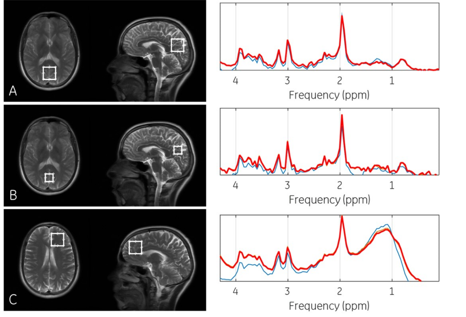
Data acquisition: SVS measurements of the brain of a healthy volunteer where acquired using a 3T-MR750w (GE Healthcare, Milwaukee, WI) with a 12-channel head receive coil. Three voxels were acquired: (SV1) Occipital lobe with size=30x30x30mm3, (SV2) Occipital lobe with size=20x20x20mm3 and (SV3) Right-Frontal lobe with size=30x30x30mm3. The acquisition parameters were: TE/TR=35/1000ms, spectral bandwidth=2kHz. PRESS volume localization was used to acquire a standard 64-average SVS and an MRSI grid with 8 phase encodings in the SI and AP directions without changing the PRESS voxel nominal resolution. Additionally, high-resolution B0-field map and coil sensitivity maps were also obtained.
Data Processing: The MRSI data was reconstructed with an overdiscrete factor of $$$\zeta$$$=5 and Gaussian target function with $$$\sigma$$$=3 sub-voxels for SRF optimization. Finally, standard SVS spectra were coherently averaged and reconstructed using standard Fourier transform. Local B0-field maps and coil sensitivity profiles were extracted using the voxel location and size.
Results and Discussion
The simulation results demonstrate the noise level reduction and the improved linewidth of the metabolite peaks. A potential 2-3-fold improvement in SNR that can be achieved using receiver coils with increased number of elements. Moreover, it is demonstrated that the line shape of the simulated metabolite peak remains unaffected after reconstruction. Fig.-4 show the in-vivo reconstruction results were a reduction of spectral artifacts were observed and a factor of 1.6 SNR improvement was achieved, corresponding to a reduction factor of 3-4 compared to regular averaging acquisitions. Lipid contamination in SV3 in Fig.-4A, show a just slight decrease. To achieve a better the suppression of nuisance signals, a higher resolution phase encoding matrix might needs to be acquired to obtain a sub-voxel spatial distribution of the excited tissues.Conclusion
Here we presented that an improvement in SNR can be achieved for SVS by using spatial encoding and an overdiscrete reconstruction, allowing to detect metabolites more reliably. Alternatively, it is possible to reduce voxel size by optimizing the SRF, hence improving spatial localization and reducing partial volume effects. Moreover, reduced voxel sizes and correction of inhomogeneities can enable acquisitions in challenging brain areas.Acknowledgements
No acknowledgement found.References
[1] Hurd R. Volume Spectroscopy Having Image Artifact Reduction. US Patent No. 5,804,966.
[2] Pruessmann K and Tsao J. Magnetic Resonance Imaging Method. US Patent No. 7,342,39.
[3] Kirchner et al. Reduction of voxel bleeding in highly accelerated parallel 1H MRSI by direct control of the spatial response function. Magn Reson Med 2014.
[4] Kirchner et al. Mechanisms of SNR and Line Shape Improvement by B0 Correction in Overdiscrete MRSI Reconstruction. Magn Reson Med 2015.
Figures