5500
Highly resolved 1H decoupled spectroscopy with a shorter constant time delay using the 2D CT-PRESS with J refocusing1Center for Environmental Measurement and Analysis, National Institute for Environmental Studies, Tsukuba, Japan
Synopsis
The 2D constant time localized sequence of CT-PRESS with J refocusing was proposed, having features of good peak resolution via 1H decoupling along F1 and of J refocusing by a 90-degree pulse at the first echo time. By controlling the time point of J refocusing, shorter constant time delay of Tct can be achieved. This method was developed on a 4.7 T whole-body MR system. Phantom experiments were demonstrated using brain mixture solutions. Three resonances at 2.28 ppm of GABA C2H, 2.35 ppm of glutamate C4H and 2.44 ppm of glutamine C4H were resolved on the phantom spectra.
Introduction
While glutamate (Glu) and γ-aminobutyric acid (GABA) are major neurotransmitters in human brain, glutamine (Gln) is a precursor and storage form of Glu, which plays an important role in the Glu-Gln cycle. Since the concentrations of these metabolites may give us the useful information, accurate quantitation is expected in 1H MRS. However, these peaks are heavily overlapped on the conventional 1H spectra due to strong couplings with small chemical shift difference and JHH coupling. This may lead to difficulty in the quantitation. To overcome this problem, we have reported in vivo detection of Glu, GABA and Gln in the human brain using two kinds of 2D localized constant time (CT) methods of CT-COSY1 and CT-PRESS2. These CT methods have a feature of the better peak resolution by 1H decoupling along the F1 direction. However, the CT methods required a long CT delay (Tct) of around 110 ms2 to minimize the anti-phased J coupled spins. This causes T2 decay and quantitation error occurs. In this work, we applied the technique3-5 of 1D spin echo with J refocusing to the CT method and proposed the modified CT-PRESS with J refocusing. A shorter CT delay is expected by controlling the time point of J refocusing. We developed the proposed sequence with Tct of 38 ms on our 4.7T MR system and demonstrated phantom experiments.Methods
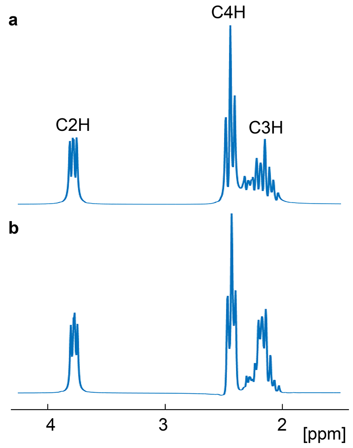
The SE sequence with J refocusing consists of 90°x-[τ-180°y-τ-90°y-τ-180°y-τ]n-acquisition where n denotes the number of cycles3-5. J is refocused by the 90°y pulse at each echo time of 2(2n-1)τ. Although a shorter delay of τ around 5ms should be set, a longer delay may be adapted for a localized CT method on whole-body scanners. Figure 1a shows a simulated glutamate spectrum at 4.7 T with τ of 12 ms, resulting in the J refocusing point of 48 ms. All glutamate peaks are almost refocused. Figure 1b shows the other by 90°x-TE1/2-180°y-TE1/2-90°y-TE2/2-180°y-TE2/2-acquisition with TE1 of 24 ms and TE2 of 14 ms. Although the total TE of 38 ms is shifted from the J refocusing point at 2TE1 of 48 ms, all peaks are relatively refocused. In the following 2D CT experiments, Tct was set to 38 ms around the J refocusing point.
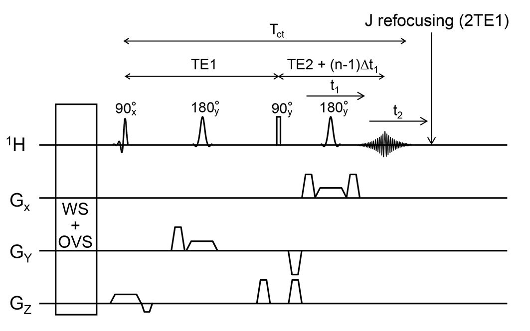
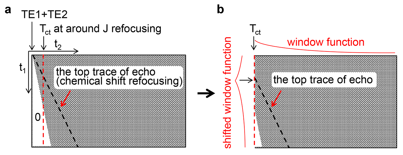
Figure 2 shows our proposed sequence of 2D CT-PRESS with a feature of J refocusing at 2TE1 by the 90°y pulse at TE1. As the second 180-degree slice pulse was shifted with Δt1/2 every t1 increment, FID was collected. After the total scan, a whole 2D time domain data were accumulated. After a suitable amount of zeros was filled in front of the acquired FIDs to meet the constant time condition2 (Fig. 3a), data were used after Tct (Fig. 3b). For weighting with this Tct, the window function shifted with the point of of echo top on the Tct line was applied along t1 (Fig. 3b). After applying window functions along t2, zero-filling and 2D FT, the CT-PRESS spectrum was obtained.
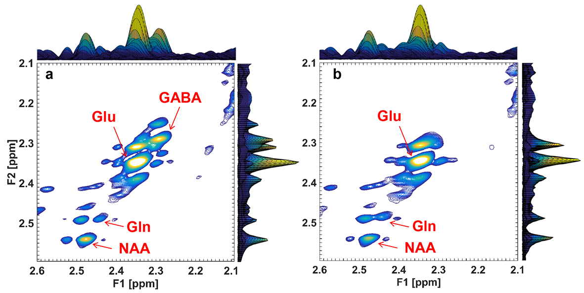
All experiments were performed using a 4.7 T whole-body MR system (INOVA, Agilent). A volume TEM coil was used both for transmission and reception. We measured spectra of CT-PRESS with J refocusing of two kinds of phantoms: the one contains a brain metabolite mixture of 10.1-mM NAA, 7.9-mM Cr, 9.0-mM Glu, 2.0-mM GABA and 3.0-mM Gln and the other that mixture without GABA. A 200-mL bottle containing each solution was placed in a saline bath and each CT-PRESS signal was acquired inside an 8-ml voxel within that bottle with a measurement time of 20 min. In all measurements, TE1 = 24 ms, TE2 = 8 ms and Tct = 38 ms. Spectral widths along F1 was 1 kHz and that along F2 was 2 kHz. The total number of t1 increments was 150 and that of complex data points in t2 was 4096. The number of average was 2. Relaxation delay was 4 s.
Results & Discussion
While three resonances at GABA C2H, Glu C4H and Gln C4H were resolved on the spectra from the solution with GABA (Fig. 4a), none arose at around 2.28 ppm without GABA (Fig. 4b). Then, it was confirmed that three resonances of Glu, GABA and Gln could be resolved on the CT-PRESS spectra with J refocusing with Tct of 38 ms. Tilted peaks may be minimized by optimizing window function.Conclusions
Our proposed method of CT-PRESS with J refocusing is useful for resolving peaks of strongly coupled spins, such as glutamate, GABA and glutamine with moderate Tct of 38 ms. This technique will be helpful to achieve accurate quantitation with further shorter Tct.Acknowledgements
No acknowledgement found.References
1. Watanabe H, Takaya N, Mitsumori F. Simultaneous observation of glutamate, g-amino butyric acid and glutamine in human brain at 4.7T using localized two dimensional constant-time correlation spectroscopy. NMR in Biomed. 2008;21(5):518-526.
2. Watanabe H, Takaya N, Mitsumori F. Highly resolved 1H spectroscopy of the human brain using ISIS CT-PRESS with resolution enhancement. Magn. Reson. Med. Sci. 2012;11(4):235-241.
3. Pan J W, Avdievich N, Hetherington H P. J-refocused coherence transfer spectroscopic imaging at 7 T in human brain. 2010;64(5):1237-1246.
4. Aguilar J. A., Nilsson M., Bodenhausen G., Morris G. A. Spin echo NMR spectra without J modulation. Chem. Commun. 2012;48:811-813.
5. Lin Y., Lin L, Wei A, Zhong J, Chen Z. Localized one-dimensional single voxel magnetic resonance spectroscopy without J coupling modulations. 2015;early view
Figures