5496
Regional GABA concentration comparison in the human brain with the interleaved short TE sLASER and MEGA-sLASER sequence at 7T1Erwin L. Hahn Institute for Magnetic Resonance Imaging, University of Duisburg-Essen, Essen, Germany, 2Donders Institute for Brain, Cognition and Behavior, Radboud University, Nijmegen, Netherlands
Synopsis
GABA is challenging to resolve due to j-couplings and overlapping signals. Previously proposed GABA editing ans short TE approach at UHF make it possible to measure relative GABA concentration. We measured GABA concentration with the interleaved sequence at various brain regions, and found an optimal method to estimate GABA in terms of spectral fitting quality. Occiptal cortex showed a high GABA concentration, and GABA editing approach gave a reliable spectral fitting quality.
Introduction
γ-Aminobutyric acid (GABA), the major inhibitory neurotransmitter, is present at circa millimolar concentrations in the human brain, and is challenging to resolve due to j-couplings and overlapping signals. Previously proposed GABA editing approach such as the MEGA semi-LASER [1] and short echo semi-LASER [2] at UHF (>7T) make it possible to measure relative GABA concentration. These two acquisition approaches have their own obvious advantages that GABA editing approach can find isolated clear GABA at 3ppm, and the short echo time (TE) approach allows the detection of an increased number of small metabolites and has a SNR advantage over long TE. Previously, Terpstra et al [3] reported approximately 11% of GABA/CR concentration ratio by MEGA-PRESS at 7T. This study aims to compare GABA concentration at various brain regions with different acquisition approaches by the interleaved sequence, test whether this approach can find the GABA concentration level reported by Terpstra et al, and find an optimal method to estimate GABA in terms of spectral fitting quality.Material and Methods
Short TE sLASER (TE/TR/NEX = 38ms/4500ms/64) and MEGA-sLASER (TE/TR/NEX = 80ms/4500ms/32) spectra were acquired using a single interleaved sequence with a voxel size of 20x20x20mm3 using a 7T system (Siemens, Erlangen), equipped with a 32Ch head coil (NOVA, NY). This interleaved sequence (Fig.1), which we combined two sequences into one single sequence to minimize factors such as motion, involves the sequential four repetition blocks of MEGA ON, MEGA OFF and two Short TEs with 32 averages per block. Total acquisition time was 9min 54sec. Six MRS spectra were acquired from six different brain regions (anterior cingulate cortex (AC), dorsolateral prefrontal cortex (DLPFC), mortor cortex(MC), occipital cortex(OCC), posterior cingulate cortex (PC), and precuneus (PrC)) on 15 healthy volunteers (12M/5F;29.34±4.49YO). As an anatomical reference, a 3D MPRAGE was also acquired before spectroscopy. B0 shimming was performed by FASTESTMAP[4]. NMRSIM (Bruker, Rheinstetten) simulated spectral fitting models with the same sequence and scan parameters of the in-vivo spectroscopy scan. LCModel estimated a GABA/tNAA concentration ratio and a Cramér–Rao lower bound (CRLB) [5], which is an estimated error of the metabolite quantification (expressed in %SD).Results
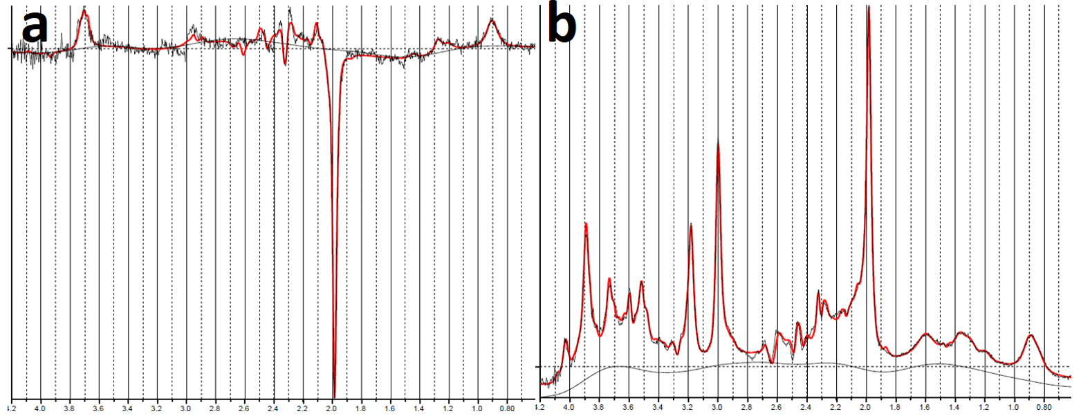
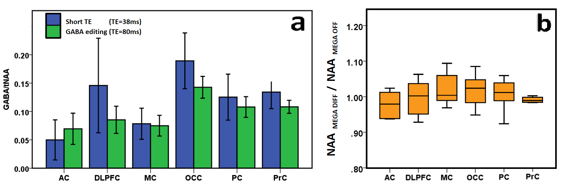
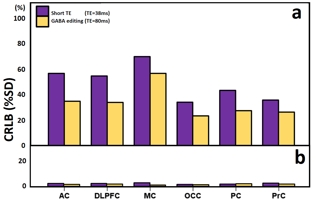
Fig.2 shows spectral fitting results of the Short TE sLASER(a) and the MEGA-sLASER(b) by LCModel. Fig 3 (a) shows a result of a GABA/tNAA concentration ratio comparison between the short TE sLASER and MEGA-sLASER for six brain regions. We could find a relatively high GABA/tNAA ratio at OC (0.188±0.038, 0.014±0.016), and a relatively low ratio at AC (0.059±0.023, 0.069±0.026) as compared with other four regions where GABA/tNAA ratio was similar (DLPFC: 0.059±0.023, 0.069±0.0262, MC: 0.078±0.031, 0.075±0.020, PC: 0.125±0.045, 0.107±0.020, and PrC: 0.134±0.0292, 0.108±0.011; Short TE sLASER, MEGA-sLASER; respectively). Similar NAA signal intensity of MEGA OFF and MEGA DIFF spectrum confirmed a stable efficiency of the MEGA inversion pulse (Fig. 3(b)). The GABA editing approach also showed better spectral fitting quality according to CRLB than the short TE approach for all regions examined (Fig. 4).Discussion
This study compared GABA concentration for six different brain regions with the short TE approach and the GABA editing approach by one single interleaved sequence acquisition. This simultaneous acquisition of short TE sLASER and MEGA-sLASER minimizes the effect of motion in the resulting spectra. GABA concentration of the short TE approach showed higher than that of GABA-editing approach. However, GABA-editing approach was superior in spectral fitting quality to the short TE approach. GABA-editing method by MEGA-sLASER is more efficient for finding a metabolite in isolated peaks than the short TE method, which need to find a small metabolite from a mixed signal. MEGA-sLASER enables accurate GABA detection.Acknowledgements
No acknowledgement found.References
[1] Andreychenko, Anna, et al. "Efficient spectral editing at 7 T: GABA detection with MEGA-sLASER." Magnetic resonance in medicine 68.4 (2012): 1018-1025.
[2] Scheenen, Tom WJ, et al. "Short echo time 1H-MRSI of the human brain at 3T with minimal chemical shift displacement errors using adiabatic refocusing pulses." Magnetic resonance in medicine 59.1 (2008): 1-6.
[3] Terpstra, Melissa, Kamil Ugurbil, and Rolf Gruetter. "Direct in vivo measurement of human cerebral GABA concentration using MEGA-editing at 7 Tesla." Magnetic resonance in medicine 47.5 (2002): 1009-1012.
[4] Gruetter, Rolf, and Ivan Tkác. "Field mapping without reference scan using asymmetric echo-planar techniques." Magnetic resonance in medicine 43.2 (2000): 319-323.
[5] Provencher, Stephen W. "Estimation of metabolite concentrations from localized in vivo proton NMR spectra." Magnetic resonance in medicine 30.6 (1993): 672-679.
Figures