5482
Phase-rotation acquisition to study imperfections of PRESS1Institute for Biomedical Engineering, University and ETH Zurich, Zurich, Switzerland
Synopsis
A phase-rotation acquisition scheme is able to separate different signals in PRESS. In this work phase-rotation PRESS measurements were complemented by simulations to assess origins of spectral distortions in cardiac spectroscopy. Simulations show that, when using a phase-rotation acquisition scheme for PRESS without spoilers, more motion is acceptable when pulses are better calibrated. However, because of non-ideal pulse profiles, spoilers cannot be fully omitted if moving tissue is studied. It is shown that phase-rotation acqusition can, by combining simulations and measurements, be used as an elegant tool to investigate spectral distortions and optimize sequence parameters in PRESS of moving tissue.
Introduction
In point-resolved spectroscopy (PRESS), spurious signals are suppressed by gradient spoiler pairs placed around each of the 180° refocussing pulses. These gradient pairs are sensitive to first and higher orders of motion, which can lead to motion-induced signal loss, especially in cardiac spectroscopy1. To overcome these effects, spoilers can be motion-compensated2 at the expense of prolonged echo times.
Imperfect power calibration has recently been studied experimentally and was found to be another major determinant of signal loss in cardiac spectroscopy3.
Originally proposed as a spoiler-free approach to spectral localization, the principle of phase-rotation4 acquistion is proposed herein to study imperfections and to optimize PRESS sequences for applications in moving tissue. In the present work in-vivo phase-rotation PRESS measurements are complemented by simulations to assess origins of spectral distortions in cardiac spectroscopy.
Theory
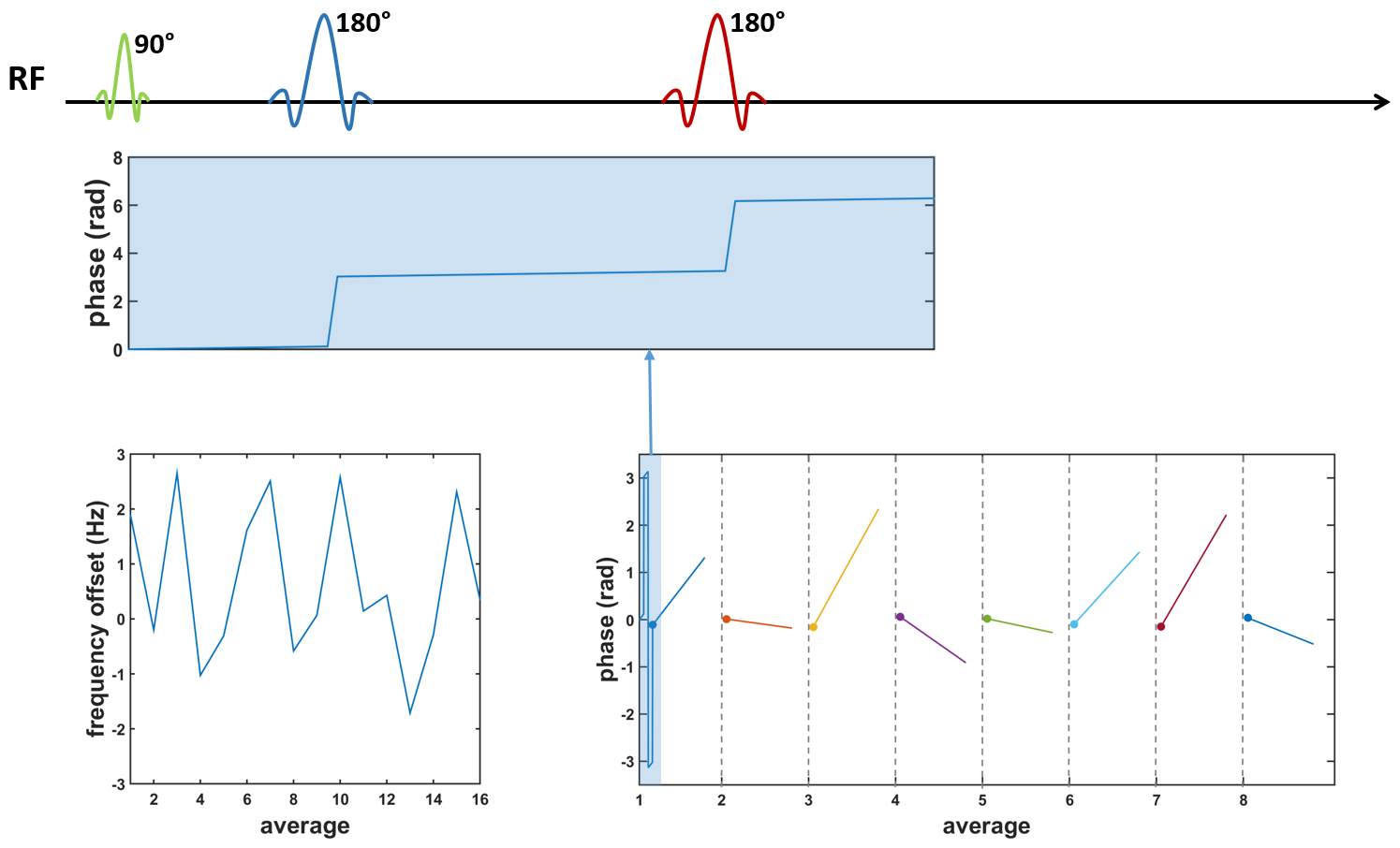
The phase-rotation acquisition scheme4 for PRESS is based on constant phase increments Δϕ1, Δϕ2 and Δϕ3 for each of the three RF pulses from one acquisition to the next. Time-domain signals obtained with each acquisition are read into a 2D matrix, which is subsequently 2D Fourier-transformed. FID and echo signal components are shifted along the phase dimension proportional to their respective phase offsets (Fig-1) and applied phase increments Δϕi. While the desired echo signal from all the pulses (SE123) can directly be extracted from row 14/16 in the case of 16 averages and phase angles Δϕ1=- Δϕ2=Δϕ3=22.5°, the distribution of spurious signals provides information on acquisition imperfections.Methods
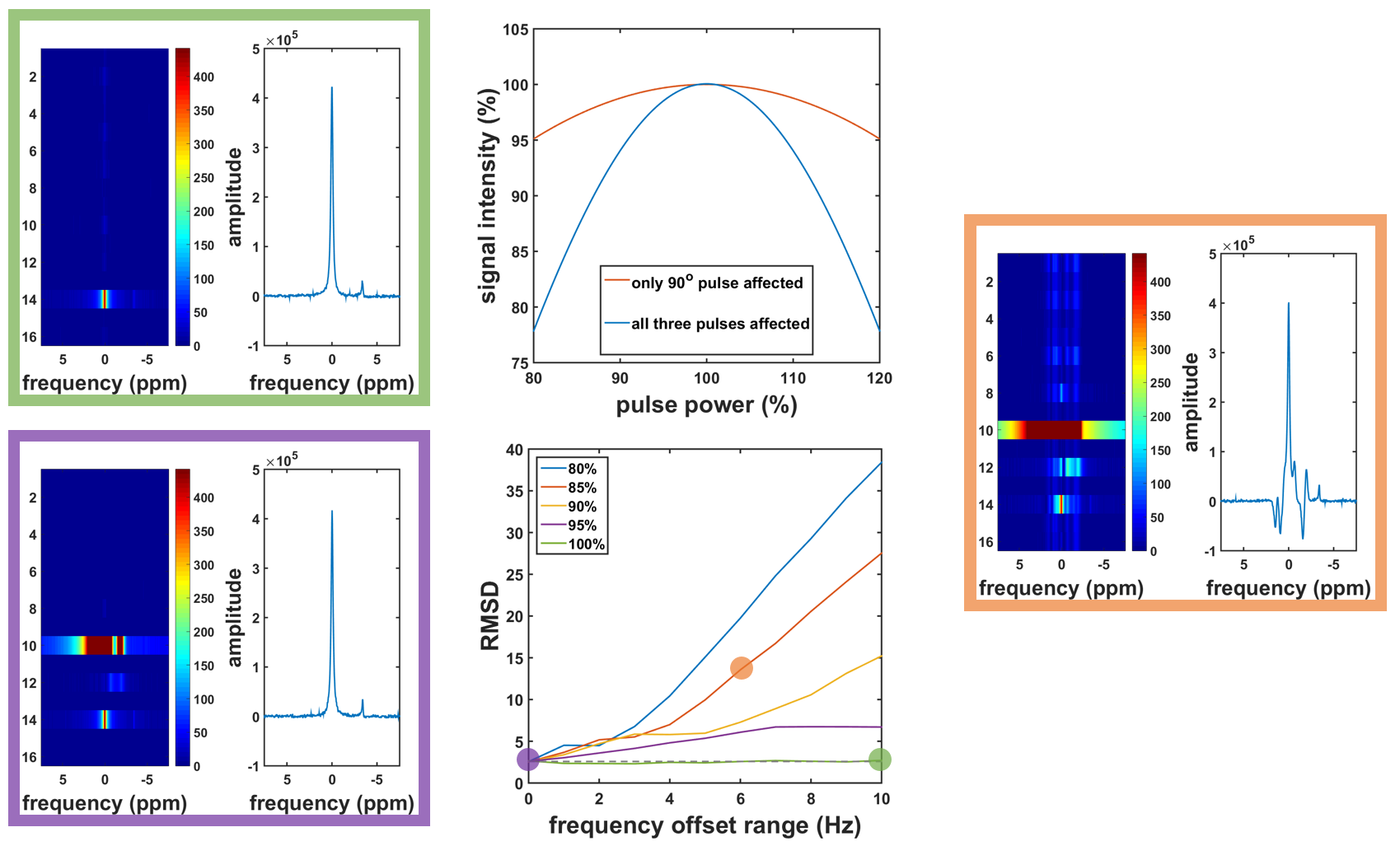
Simulations of phase-rotation PRESS were performed using MATLAB. A volume of 3x3x3 cubes was simulated in which only the center cube represents the localized PRESS voxel of interest while the other cubes are treated as surrounding tissue. The effect of imperfect power optimization was incorporated by scaling nominal flip-angles from 80% to 120%. Motion was simulated by applying random frequency offsets (range 0-10Hz) to each average to induce phase variations within one acquisition, as well as varying starting conditions for each average as illustrated in Fig-2.
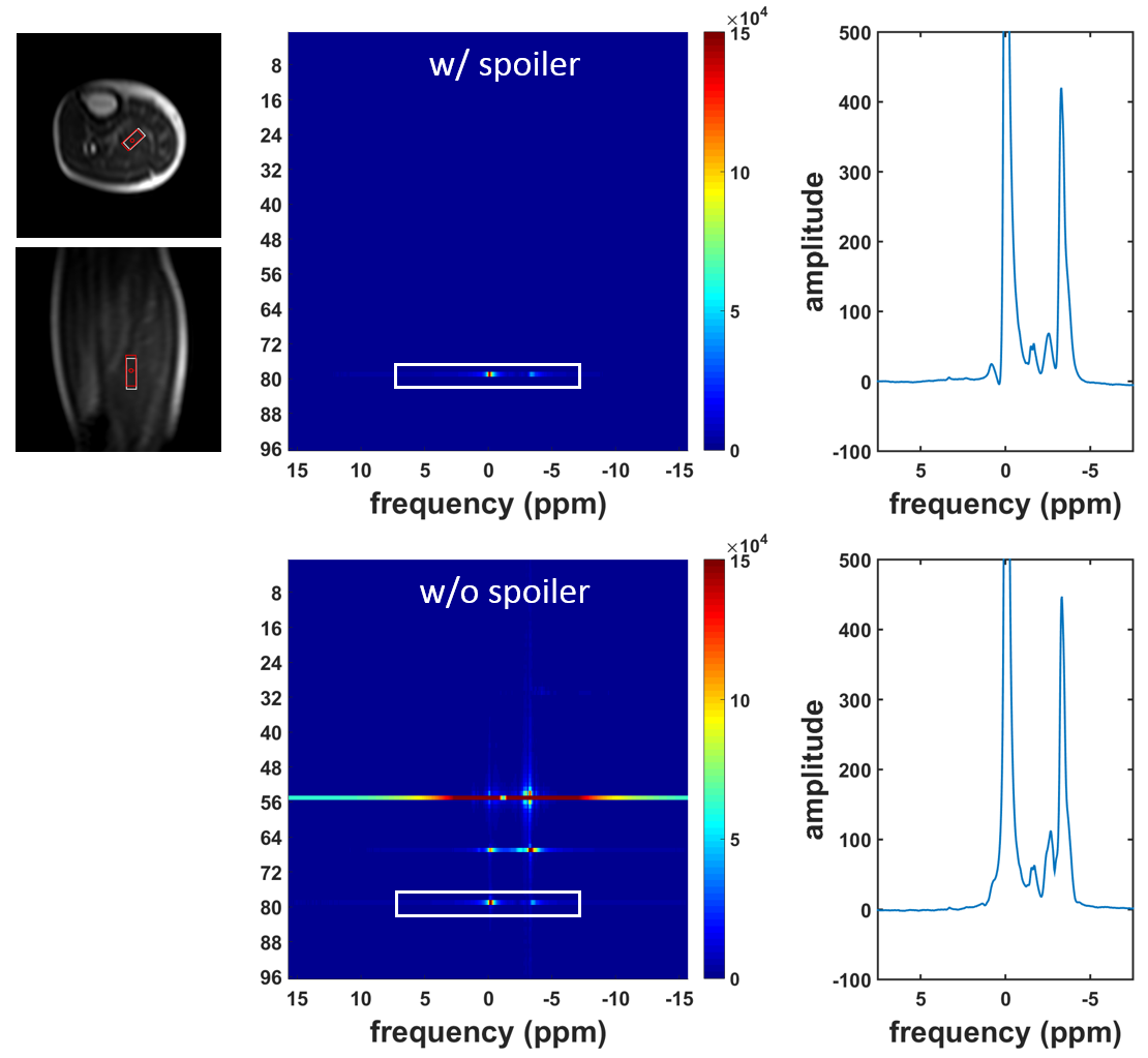
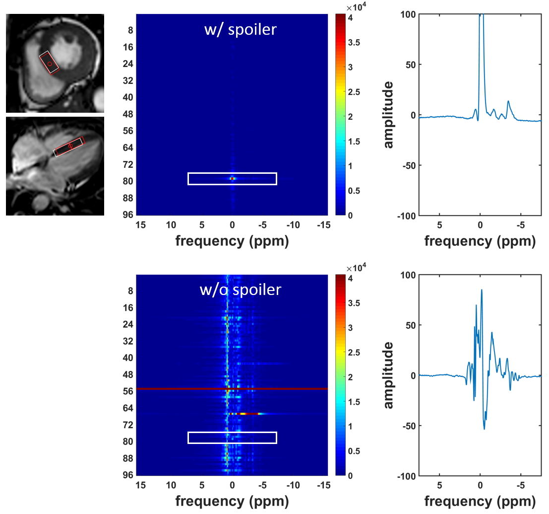
In-vivo measurements were performed on healthy volunteers upon written informed consent and according to ethics and institutional guidelines. The phase-rotation scheme was implemented on a clinical 1.5T Philips Achieva scanner, equipped with a five-channel cardiac receiver surface coil. Spectra in calf muscle and the interventricular septal wall were acquired using a PRESS sequence with reduced-spoiler areas4 as well as without spoiler gradients, with the following sequence parameters: voxel size 10×20×40mm3, spectral BW: 2000Hz, TR: 2s, TE: 20ms (with spoiling) /17ms (without spoiling), NSA: 96 (water-suppressed) /16 (water), CHESS water suppression BW 80Hz and iterative volume shimming. A respiratory navigator (4mm window) and ECG triggering (320ms, end-systole) were employed for cardiac measurements.
Phase increment values were Δϕ1=22.5°, Δϕ2=-22.5° and Δϕ3=22.5° for the 90°, first 180° and second 180° pulse, respectively.
Results
Simulated effects of power calibration and motion are shown in Fig-3. Whereas it is clear that an imperfect 90° pulse has a negative effect on signal intensity, this effect is even more pronounced if the echo-pulses are imperfect as well. To indicate the effect of motion on spectral quality for different power calibration values affecting all three pulses, spectra were simulated for different quantities of motion. The root-mean-square deviations (RMSD) of spectra with and without motion were calculated for the region between 100Hz and 300Hz. Imperfections in pulse power show up only on the phase-rotation lines as explained in Fig-1, whereas motion causes artefactual signal along the phase direction. When using a phase-rotation acquisition scheme more motion is acceptable when the pulses are better calibrated.
In-vivo results of calf muscle are shown in Fig-4. Because of a small amount of pulsatile motion in the leg, vertical dispersion can still be seen in the phase rotation diagram of the measurement without spoilers. However, spectral quality and signal amplitude are still comparable to the result of a phase-rotation experiment with spoilers. This is in contrast to phase-rotation PRESS without spoilers in the heart (Fig-5), where motion is a primary cause of spectral distortion.
Discussion
Simulations indicate that, with ideal power calibration, distortions stemming from motion can efficiently be mitigated using phase-rotation acquisitions, even in the absence of spoiler gradients. In-vivo this would, however, require homogenous B1+ over all three orthogonal excitation slices, which is not feasible. For cardiac spectroscopy spoiler gradients can therefore not be omitted, but could possibly further be optimized using phase-rotation analysis.Conclusion
Phase-rotation acqusition allows to disentangle effects of power miscalibration and tissue motion in PRESS and hence helps to optimize power settings and motion compensation strategies for cardiac singe voxel spectroscopy.Acknowledgements
No acknowledgement found.References
1. Weiss K, Summermatter S, Stoeck CT, Kozerke S. Compensation of signal loss due to cardiac motion in point-resolved spectroscopy of the heart. Magn Reson Med 2014;72:1201-1207.
2. Fuetterer M, Stoeck CT, Kozerke S. Second-order motion compensated PRESS for cardiac spectroscopy. Magn Reson Med 2016;00;00-00.
3. de Heer P, Bizino MB, Lamb HJ, Webb AG. Parameter optimization for reproducible cardiac 1 H-MR spectroscopy at 3 Tesla. J Magn Reson Imaging 2016;44(5):1151-1158.
4. Hennig J. The application of phase rotation for localized in vivo proton spectroscopy with short echo times. J Magn Reson 1992;96:40-49.
Figures
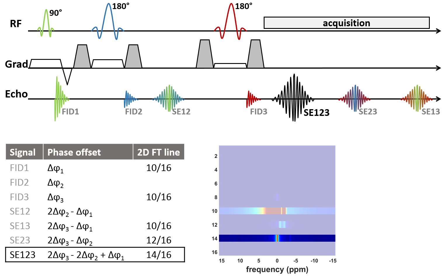
Figure 1: PRESS sequence with different signal components (top) with their corresponding phase offsets (bottom-left). In a phase-rotation experiment with 16 averages and Δϕ1=22.5°, Δϕ2=-22.5° and Δϕ3=22.5°, the proper signal appears in row 14 (bottom-right).
Note that FID signal components outside the readout window might still be observable, due to their large amplitude and relatively long T2* (diagram is not to scale).