5473
Novel Approach for Simultaneous In Vivo 31P MRS Measurements in Frontal and Occipital Lobes of Human Brain with Dynamic Shimming1Center for Magnetic Resonance Research, Department of Radiology, University of Minnesota Medical School, Minneapolis, MN, United States
Synopsis
Functional/metabolic changes in different brain regions of interest are of importance for better understanding of the pathophysiological mechanism underlying the human brain diseases. In this work, we present a novel design of the dual-channel 31P MRS system for simultaneous measurements of cerebral high-energy phosphate metabolism from two brain regions of interest by incorporating a new pulse sequence and two separated RF surface coils with a transistor-transistor logic (TTL) controller. By successfully implementing this method, we are able to obtain high quality in vivo 31P MR spectra from both frontal and occipital lobes within the same amount of time as the traditional method covering one brain region. From an engineering perspective, this new approach provides a cost-effective solution for in vivo 31P MRS study of multiple brain regions with a conventional single-channel transmitter-receiver configuration. Therefore, this valuable MR tool can be used in examining the cerebral energy metabolism across different brain regions, and the same approach could be employed to other spin applications.
Introduction
With increasing recognition of complex neurological disorders with distinct progression in different brain regions, it is essential to explore functional/metabolic impairments in various brain regions of interest; for instance, Parkinson’s disease (PD) involves motor dysfunction in motor cortex as well as non-motor cognitive deficits in the frontal lobe [1]. For this reason, a head volume RF coil has been preferred to cover the entire human brain despite high cost and a drawback of relatively lower sensitivity for detecting brain metabolites. As a cost-effective and time-efficient engineering solution, thus, we designed a novel dual-channel 31P surface coils system and incorporated that with a new pulse sequence and a transistor-transistor logic (TTL) controller for simultaneous measurements of energy metabolism from two brain regions. To test this method, the frontal and occipital lobes were selected for studying high-energy phosphate metabolism using in vivo 31P MRS in combine with the magnetization transfer (31P-MT) technique, which offers capability of examining the key bioenergetic reactions catalyzed by the creatine kinase (CK) and ATPase enzymes. [2-3]Methods
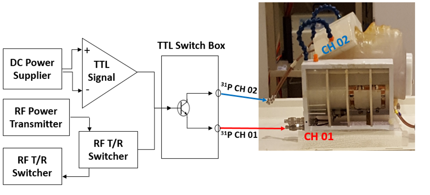
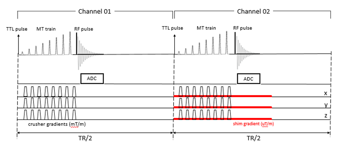
All MR experiments were performed at 7.0T/90cm whole body scanner(Siemens). Each channel targeting one brain region of interest consists of a butterfly 1H coil for anatomical imaging and B0 shimming, and a single-loop 31P coil for 31P MR spectra. The B1 insensitive selective train to obliterate signal scheme (BISTRO) [4] was applied to saturate the γ-ATP resonance followed by a single-pulse-acquire sequence (320 averaging, 3s TR, 5kHz spectral bandwidth (BW), 300 ms hard pulse with an Ernst flip angle of 84° for excitation). Moreover, 3D chemical shift imaging (CSI) was acquired using Fourier series window [5] (1.2 s TR, 5kHz BW, 300 ms hard excitation pulse, and phase encoding steps 9x9x7, FOV = 120x120x90 mm3). As shown in Fig. 1, 31P MRS data for each coil were alternatively acquired by switching each channel at the half TR and each measurement was done within the same apparent acquisition time. RF transmitter powers for 90-degree excitation pulses were separately calibrated for each channel for setup of Ernst flip angles.Results and Discussion
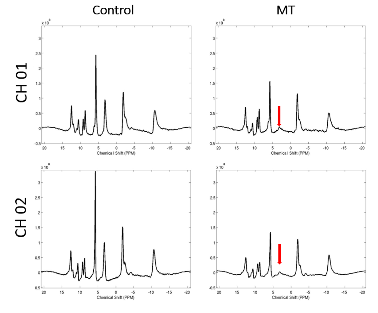
Figure 2 shows a general scheme or pulse sequence of the 31P dual-channel system for the simultaneous measurement from two targeted brain regions. Using a single transmit amplifier and receiver channel incorporating with TTL gate function, 31P MRS data were successfully collected from each channel without doubling the acquisition time. Figure 3 displays representative in vivo 31P-MT spectra of the human frontal and occipital lobes with and without γ-ATP saturation, showing excellent spectral quality (spectral linewidth of PCr < 11 Hz) and effective MT saturation efficiency. Notably, with application of additional adjustment of the linear shim gradients (see Fig. 2), the 31P MR spectrum of the frontal brain region reached a comparable quality as the occipital lobe, which enables reliable quantification of cerebral phosphorus metabolites concentration and changes in both brain regions. In addition, the 3D 31P MRS CSIs shown in Fig. 4 demonstrate the sufficient coverage and good B0 homogeneity of the dual-coil system that result in excellent spectral quality in both brain regions.Conclusion
In this work, we demonstrate a novel design of dual-channel 31P system for simultaneous measurement of the regional high-energy phosphorous metabolism in the human brains of interest. From an engineering perspective, the new approach offers a cost-effective and time-efficient solution for high quality in vivo 31P MRS measurements from multiple brain regions using a conventional single-channel transmitter-receiver configuration. Therefore, this approach could provide a valuable MR tool for examining the cerebral energy metabolism and its impairment across different brain regions. The same approach could be employed to other nuclear spin applications.Acknowledgements
NIH grants: R01 NS057560, NS070839, MH111447; R24 MH106049, S10RR026783, P41 EB015894, and P30 NS057091; and the AHC Faculty Research Development (FRD) grant from the University of Minnesota.References
[1] Taylor et al., Brain, 109: 845-883 (1986); [2] Du et al., PNAS, 105: 6409-6414 (2008); [3] Zhu et al., Neuroimage, 60: 2107-17 (2012); [4] de Graaf et al., J Magn Reson B, 113: 35-45 (1996); [5] Hendrich et al, J Magn Reson, 105:225–232 (1994).
Figures