5452
RF safety of an implanted port catheter in direct vicinity of a 7T transmit head coil1Erwin L. Hahn Institute for MRI, University Duisburg-Essen, Essen, Germany, 2MR Safety Testing Laboratory, MR:comp GmbH, Gelsenkirchen, Germany, 3Hamm-Lippstadt University of Applied Sciences, Hamm, Germany, 4Medical Physics in Radiology, German Cancer Research Center (DKFZ), Heidelberg, Germany, 5High Field and Hybrid MR Imaging, University Hospital Essen, Essen, Germany
Synopsis
Potential RF-induced heating from an implanted port catheter in direct vicinity of a local transmit head coil at 7T was investigated. The assessment included direct measurements of E-field and temperature in a rectangular head/shoulder phantom filled with tissue simulating liquid as well as numerical SAR and thermal simulations in two human body models. Two different RF coils were used for the evaluation, a custom-made 8-channel head coil and the widely-available Nova Medical head coil. No evidence of RF-induced heating was found. Identical transmit power restrictions apply with or without port for both investigated RF coil types.
Target Audience:
Radiologists and physicists interested in MR safety and ultra-high field imaging.Purpose:
Ultra-high field (UHF) magnetic resonance imaging (MRI) allows a more detailed depiction of microvascularity and microhemorrhages in tumor and ischemic diseases, high-resolution imaging of cerebral vessel pathologies as well as micro-anatomical depiction of cortical structures, i.e. aspects which offer potential benefits for patient treatment in terms of diagnostics, surgical planning, and therapy monitoring [1, 2]. On the other hand, many patients present with implanted medical devices (IMD), but only a few of these have been labeled MR conditional up to 7T magnetic field strength. The German UHF network GUFI has recently published a recommendation for the inclusion of subjects with passive IMDs, stating that safe imaging at UHF can be performed if the IMD is 3T MR conditional and outside a coil-dependent safety area [3]. This work investigates potential RF-induced heating from a port catheter typically implanted subcutaneously below the clavicula, i.e. in direct vicinity of the transmit head coil.Methods:
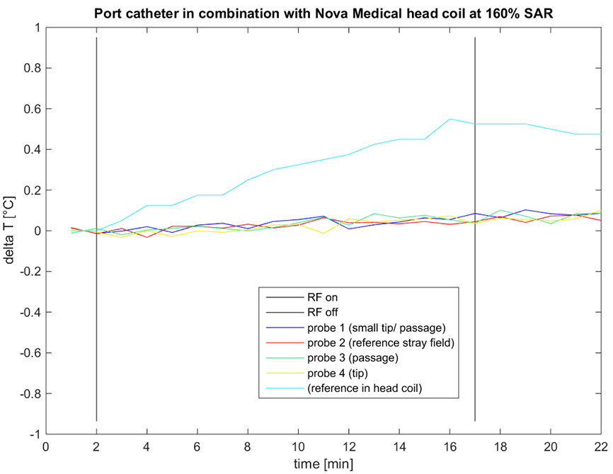
The IMD under test was a 3T MR conditional port catheter (NuPort-HP power port, PHS Medical, Germany) made of titanium (Fig. 1). The port was tested both with a custom-made 8-channel Tx/Rx head coil [4] and a commercially available 32-channel Rx/CP birdcage Tx coil (Nova Medical, NY). First, electric field measurements (ES3DV2, SPEAG, Switzerland) were compared with and without the implant present at four distances (2 cm worst-case and typical positions at 10, 13, 15 cm) with respect to the 8-channel head coil. This RF coil was selected as it could be easily integrated into the field measurement set-up (Fig. 2A/B) and allowed different driving modes via a butler matrix (CP+/CP2+). To avoid a potential drift in field measurements, the applied accepted power was set to 8.7W (at this power and for a given point, E-field values remained constant after 2 hours of exposure). For the measurements, a rectangular head/shoulder phantom filled with tissue simulating liquid was used (permittivity of 43.8 and conductivity of 0.75 S/m). Axial and coronal planes of 40 mm x 40 mm close to the port were evaluated. Second, temperature measurements according to ASTM F2182 (Fig. 2C/D) were performed with both RF coils using fiber-optic probes (Luxtron, CA). A turbo spin echo (TSE) sequence running at maximum allowed SAR (average power: 11.7W) for 21:44 min was used for heating. A TSE sequence running at 160% SAR (average power: 18.8W) for 15 minutes was additionally used for the Nova Medical head coil as a worst-case example. Here, a solution of salt and hydroxyethylcellulose (permittivity of 78.5, conductivity of 0.4S/m) was used according to the ASTM standard. Third, numerical SAR and temperature simulations (Semcad-X, SPEAG, Switzerland) for the Nova Medical head coil, performed in two heterogeneous body models (Duke and Ella [5]), concluded the RF safety assessment (Fig. 1B/C). Prior to detailed simulations the coil model was validated using B1 maps in a homogeneous phantom. Thermal simulations were performed with constant perfusion and input power levels of 25W for Ella and 22W for Duke, reaching a steady-state temperature of 39°C in the head after 60 minutes of exposure.Results:
Maximum E-fields were found within a 3 mm radius around the tip of the port and reached values of 15/8/7/6 V/m for distances between port and RF coil of 2/10/13/15 cm, respectively. Maximum percentage deviation compared to the case without port was 149% (Fig. 3). Values were similar for both CP modes. Maximum E-fields within the RF coil and without port present reached values of 30V/m and higher. Temperature measurements performed with both RF coils yielded no temperature increase at the port, nor at the contralateral side of the phantom. Also, the high SAR measurement showed no temperature increase (Fig. 4). Likewise, results from numerical simulations yielded only negligible differences in 10g-averaged SAR distributions across the port (Fig. 5 Duke). Maximum 10g-averaged local SAR remained unchanged by the presence of the port. Averaging over 1g of tissue yielded maximum SAR elevations around the port of 2.4 to 1.5 mW/g for Ella and 2.6 to 1.7 mW/g for Duke. However, the temperature values of 37.3°C remained unchanged around the port for both human models.Discussion:
Neither direct measurements of E-fields and temperature, nor numerical SAR and thermal simulations performed in two human models showed evidence of RF-induced heating due to the presence of an implanted port catheter in direct vicinity of a local transmit RF head coil at 7T. Based on these data, identical transmit power restrictions apply with or without port for both investigated RF coil types. However, a generalization for any RF coil or implant type cannot be made, and further configurations would require individual investigations.Acknowledgements
No acknowledgement found.References
[1] Kraff O, et al., MRI at 7 Tesla and above: demonstrated and potential capabilities. J Magn Reson Imaging 2015;41(1):13-33.
[2] Barrett TF, et al., Application of ultrahigh field magnetic resonance imaging in the treatment of brain tumors: a meta-analysis. World Neurosurg. 2016 Feb; 86:450-65.
[3] GUFI recommendation on “Approval of subjects for measurements at ultra-high-field MRI”, version 01/21.01.2016, http://www.mr-gufi.de/index.php/en/documents
[4] Orzada S, et al., 8-Channel Transmit/receive Head Coil for 7T Human Imaging Using Intrinsically Decoupled Strip Line Elements with Meanders. Proceedings ISMRM, 2009, abstract 3010.
[5] Christ A, et al., The Virtual Family--development of surface-based anatomical models of two adults and two children for dosimetric simulations. Phys Med Biol 2010; 55(2):N23-38.
Figures
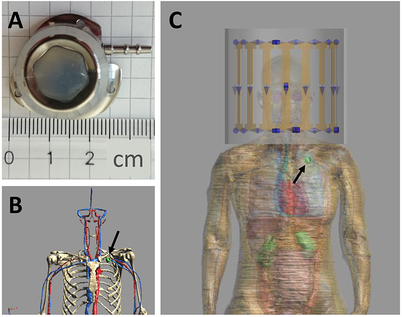

Figure 2: A: Set-up used for E-field measurements and enlarged view of field probe and port. B: Schematics of port placement and measurement planes (green) within rectangular phantom. Red dashed line indicates RF coil. C: Set-up used for fiber-optic temperature measurements with Nova Medical head coil. D: Enlarged view of port and probe placements.