5451
High Resolution Volume Of Interest Acquisition With 3D Gradient Echo Sequence Using Reduced FOV 2D-RF Excitation Pulse.1MRI Research, University Children Hospital, Zurich, Switzerland, 2BioFlow, University Picardie Jules Verne, Amiens, France
Synopsis
We designed high spatio-temporal resolution imaging sequence by reducing the excited 3D volume to the object of interest in the slab. A fast spoiled gradient recalled echo acquisition sequence was modified by incorporating spiral selective 2DRF excitation in 2 perpendicular gradients. Three different combinations of slice-select gradient axis (XY, YZ, and XZ) were tested simultaneously in axial, coronal and sagittal prescription planes and different imaging parameters to define the best scheme that provides extremely high resolution spatial imaging, with low SAR and minimum artefact on clinically available scanner.
Purposes
Gradient-recalled echo sequences provide high spatial resolution images because they are suited for 3D anatomical imaging. Different techniques have been suggested to increase the in-plane spatial resolution, but none was applying 2D RF excitation pulses [1,2]. This multidimensional selective method has been widely used in 2D imaging such saturation and inversion [3] and was validated for brain DTI [4], abdominal fat-water separation [5] and cardiac black-blood MRI [6]. Previous study suggested the use of 2D spiral gradients with RF excitation in a 3D fast gradient recalled echo sequence [7], but did not compare the different plane prescription (axial, coronal, and sagittal) and the different combination gradient waveforms (XY, YZ and XZ axes) that provide higher imaging quality. The purpose of this study was to extend the use of 2DRF to true 3D volume imaging acquisition with fast spoiler gradient recalled acquisition (FSPGR) and to compare the plan prescription effect and the slice selection gradients combination on volume rendering and slice profile.Material and Methods
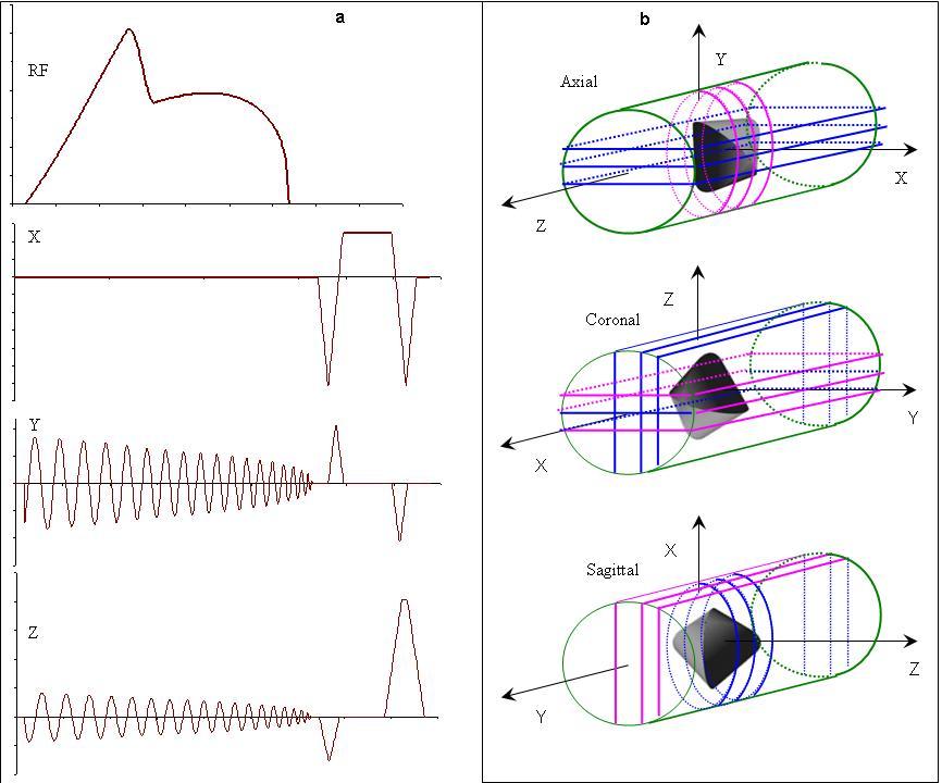
A commercial steady-state free precession sequence (fast imaging employing steady state acquisition, FIESTA) was modified by incorporating a two dimensional spiral excitation pulse with 2 oscillating gradients. The RF pulse is a half-Gaussian with a 6ms duration and the 2 oscillating gradients are designed like inward spiral with trajectories ending at the center of k-space and with no need to any rephasing pulse. The spatial selective gradient waveforms are similar to that used for spiral readout gradient in echo-planar imaging to fill the k-space. Three different schemes designed 2 perpendicular axes are: (X and Y), (Y and Z), and (X and Z=. The diameter of the excited cylindrical volume (i.e. thickness of the 2D profile) is given by the following equation D = (T.Δf)/(2.Kr) T.Δf = time bandwidth product and Kr = Maximal k-space radius.
Image aliasing occurs when the Nyquist theorem
applied in radial direction Δkr = 1/D is violated (D is the diameter of the cylindrical
FOV) and Δkr is the distance between 2 k-space points in the radial
direction the spatial radius of the cylinder r > 1/Δkr. A cylindrical phantom was chosen for this experiment (Figure 1). The sequence was
tested on 3T scanner (GE healthcare,
gradient amplitude = 40 mT/m,
slew-rate = 200 T/m/s) using 8-channel torso coil (Figure 1). The imaging
parameters were as follow: flip angle = 12°,
TE = min full, TR=12s, 2 Nex, matrix
= (128,
96), image reconstructed in (256,
256), FOV=96 mm2, phase FOV=100%,
slice thickness = 1.2 mm (zipped by factor of 2),
BW=31.25 kHz. The images were acquired in 3 different planes (axial, coronal, and sagittal) for each of the 3 gradient waveforms combinations while the imaging parameters were all equal.
Results
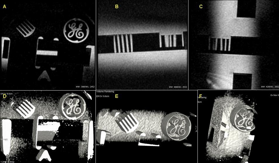
The suggested sequence has low SAR because the calculated peak B1 = 0.003238 G, the maximum integral B12 = 0.0002926 G2, and the energy deposited = 0.007955 J at 12° flip angle. A slab volume of 32 slices can be acquired in 90 seconds (or less when using fractional FOV on Y axis). The experiments clearly show that the gradient waveforms combination of Y and Z axes provides the best image quality in term of high signal intensity and fewer image artifact. This has been proven for 2D display and 3D image reconstruction (Figure 2). These results were validated in any plane and reproducibility was has been proved fro any imaging parameters. The 2 other gradient waveform combinations (XY or XZ) showed more artefacts (aliasing, phase shift)Discussion
We have demonstrated that extremely high resolution 3D images are made possible with 2D RF excitation and slice select oscillating gradient waveforms on Z and Y axes with gradient recalled sequence. The suggested sequence had low SAR, high signal intensity, sub-millimeter slice thickness (0.6 mm) and in plane resolution < 0.37 mm2 . The reproducibility has been shown in axial, coronal and sagittal planes. This modification can be designed in any 3T or 1.5T clinical scanner and thus might be used in clinical applications. Future works include decreasing the acquisition bandwidth (Hz/pixel) to increase SNR and the use of frequency modulation pulse [8].Acknowledgements
No acknowledgement found.References
[1] Feinberg et al., Radiology (1985);
[2] Pauly et al., JMR (1987);
[3] Alley et al. MRM (1997);
[4] Finterbusch, ISMRM 17, 165 (2009);
[5] Yuan et al., ISMRM 17, 2776 (2009);
[6] Abd-Elmoniem et al., MRM (2012);
[7] Makki M, ISMRM 23, 1866 (2015) ;
[8] A Jang et al., MRM 76 (2016)Figures