5440
Percutaneous MRI-guided focal cryoablation of recurrent prostate cancer: how we do itChristiaan G. Overduin1, Joyce G.R. Bomers1, Michiel J.P.M. Sedelaar2, Sjoerd F.M. Jenniskens1, and Jurgen J. Fütterer1,3
1Radiology and Nuclear Medicine, Radboud University Medical Centre, Nijmegen, Netherlands, 2Urology, Radboud University Medical Centre, Nijmegen, Netherlands, 3MIRA Institute for Biomedical Engineering and Technical Medicine, University of Twente, Enschede, Netherlands
Synopsis
We present a feasible and safe approach to perform transperineal MRI-guided focal cryoablation in patients with recurrent PCa after radiotherapy, with encouraging initial results.
Purpose
To describe our approach to transperineal MRI-guided focal cryoablation of recurrent prostate cancer (PCa).Outline of content
From May 2011 to November 2016, 65 consecutive patients with PCa recurrence after radiotherapy underwent transperineal MRI-guided focal cryoablation at our institution. Patients were treated under general anesthesia. Procedures were performed in a 1.5T or 3T MR system (Magnetom Avanto/Skyra, Siemens, Erlangen, Germany). In this educational exhibit, we describe step-by-step how we perform this interventional procedure. Technical details on procedure setup, equipment, typical procedure parameters and used MRI sequences will be provided. Imaging examples will be presented. MR imaging findings observed during needle placement and cryoablation monitoring will be reviewed (Figure 1). Diagnostic work-up and follow-up imaging protocols are discussed. Finally, we report our initial clinical experience with this treatment.Summary
We present a feasible and safe approach to perform transperineal MRI-guided focal cryoablation in patients with recurrent PCa after radiotherapy, with encouraging initial results.Acknowledgements
No acknowledgement found.References
No reference found.Figures
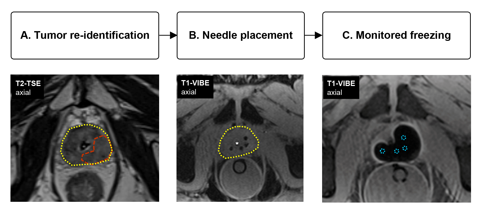
Figure 1 – Overview of main
procedure steps of MRI-guided focal prostate cryoablation. A) T2-weighted turbo
spin echo (TSE) imaging of the prostate (yellow outline) is used to re-identify a biopsy-proven local PCa recurrence in the left peripheral zone (orange
outline). B) T1-weighted volume interpolated gradient echo (VIBE) imaging shows
placement of four cryoneedles in the prostate near the target region. A
urethral warming catheter has been introduced (*). C) Freezing
is applied under continuous monitoring with T1-weighted VIBE imaging; Image
shows final frozen area at the end of the second freeze cycle.